Preparing for stoma reconstruction surgery. Colostomy closure - reconstructive coloplasty. Potential complications of a colostomy
At the present stage of surgical treatment of various diseases of the large intestine, there are certain successes, however, often it is still necessary to complete a radical operation with the imposition of an unnatural anus, or colostomy.
The presence of a colostomy on the anterior abdominal wall, often (in 25-45% of cases) complicated, disables patients, causing them severe physical and mental suffering. Therefore, the restoration of the continuity of the colon is of decisive importance for the social and labor rehabilitation of this contingent of patients, this allows them to return to active work.
However, the results of reconstructive operations after closing even such seemingly simple types of colostomy as parietal, loop and double-barrel colostomy on a separate loop cannot satisfy coloproctologists at present. So, according to Vorobyov G.I. et al. (1991), Salamova K.N. et al. (2001), Kunin N. et al., (1992), Parker S . L. et al. (1997) the frequency of wound suppuration reaches 35-50%, anastomosis suture failure with fistula formation - 20-23%, and in some cases these operations lead to deaths, which are 1-4%.
In patients with a single-barrel end colostomy after Hartmann's operation, plastic surgery, which is more complex in clinical practice, is required to restore the continuity of the large intestine. There are reports in the literature on this issue (Khanevich M.D. et al., 1998; Trapeznikov N.N., Axel E.M., 1997; Flue M. et al., 1997).
All this indicates the relevance of this problem and necessitates scientific research to improve the results of restorative operations in patients with various types of colostomy.
During the period from 1993 to 2003, the department of coloproctology restored the continuity of the colon in 283 patients with various types of colostomy. There were 176 men and 107 women. The age of patients is from 18 to 70 years.
Indications for colostomy were various diseases and injuries of the colon. The largest group consisted of 213 patients with malignant tumors of the colon (table 1). At the same time, parietal and loop colostomy were formed in 63 patients (Group 1), double-barreled separate colostomy after resection of a segment of the colon - in 73 (Group 2), single-barreled (terminal) colostomy after Hartmann's operation - in 147 patients (3 i group).
Table 1. The nature of the disease and the type of formed colostomy
| Past illnesses | Type of colostomy |
Total patients % | ||
Wall and loop |
Double-barreled separate | single barrel | ||
colon cancer |
||||
Colon injury |
||||
Complicated |
||||
colonic diverticulosis UC and disease |
||||
Crown thick |
||||
| guts | 1 | 6 | 4 | 11 (3,9) |
The recovery time of intestinal patency ranged from 1 month. up to 4 years after the imposition of a colostomy and depended on the general condition of the patients, the absence of a recurrence of the disease, the presence of pericolostomy complications and inflammatory processes in the abdominal cavity.
In preparing the colon for surgery without using antibiotics, special attention was paid to the mechanical cleaning of the adductor colon and adaptation of the disconnected segment. Regardless of the method of closure and type of colostomy during the operation, great importance was attached to the primary suturing of the stoma and the sequence of stages of performing a reconstructive operation.
To ensure recovery operations in patients with various types of colostomy, we used epidural anesthesia, prolonged in the postoperative period, in combination with intravenous intraoperative infusion of Diprivan or Calipsol. It contributes to the early awakening of patients, provides adequate pain relief, normalizes the motility of the organs of the gastrointestinal tract, thereby creating favorable conditions for the healing of reconstructive anastomosis. Against the background of rational preoperative preparation and pathogenetically substantiated therapy, it ensures a smooth course of the operation and the postoperative period.
When performing a reconstructive operation in patients of the 1st and 2nd groups, both extraperitoneal and intra-abdominal methods of stoma closure were used. The results of these operations were as follows. Of the 136 operated groups, the postoperative period proceeded smoothly in 113. In 23 patients, various complications were observed, mainly suppuration of the abdominal wall wound at the site of the former colostomy - in 18 patients (13.2%) and failure of the anastomotic sutures with the formation of a colonic fistula - in 5 ( 3.7%. At the same time, in all patients, the fistulas subsequently closed after conservative treatment. There were no lethal outcomes in the groups under consideration.
When analyzing the frequency and nature of postoperative complications, depending on the method of closing the colostomy, we found an undeniable advantage of the intra-abdominal method of closing the stoma compared to the extra-abdominal one (Table 2).
Tab. 2 Colostomy closure methods and types of complications
Colostomy closure method |
Total patients |
Complications |
|
suppuration |
|||
| Extraperitoneal | 8 (26,9%) | ||
| intraperitoneal | 63 | 5 (7,9%) | 1 (1,6%) |
| TOTAL: | 13 (14,4%) | ||
So, with the extraperitoneal closure method, wound suppuration was observed in 22.2%, and suture failure - in 11.1% of the operated patients. Despite the fact that the intraperitoneal method was more often used in patients with various pericolostomy complications, wound suppuration in the postoperative period was only in 10% of patients, i.e. occurred almost 3 times less often, and the failure of the anastomotic sutures with the formation of a fistula - in 1%, or more 10 times less often compared with the extraperitoneal closure method. The extraperitoneal method of closing the colostomy can be recommended for the treatment of patients with parietal or loop colostomy in the presence of a flat pliable spur and the absence of pronounced cicatricial changes in the tissues surrounding the stoma.
In patients with a single-barrel (terminal) colostomy who have undergone a Hartmann-type bowel resection, plastic reconstructive surgery is usually required to restore colonic continuity. The complexity of such an operation is due to the severity of the cicatricial adhesive process in the abdominal cavity, small pelvis, sometimes significant diastasis of the colon segments, as well as the presence of a short rectal stump located under the pelvic peritoneum.
The optimal period of reconstructive surgery in patients of this group should be considered 6-12 months. after radical surgery. This period is necessary to restore the patient's strength after the first operation and eliminate inflammatory processes in the abdominal cavity and small pelvis, which often accompany radical operations.
The choice of a coloplastic method for restoring the continuity of the large intestine after the Hartmann operation depended on the length of the disconnected intestine and the severity of the inflammatory process in the small pelvis. With proper mobilization of the colon, it is almost always possible to perform a more physiological and less traumatic coloplasty.
The most formidable complication after reconstructive surgery is anastomotic suture failure, which we observed in 12 patients (8.2%). At the same time, emergency relaparotomy, drainage of the abdominal cavity and the formation of a proximal colostomy were undertaken to treat the resulting peritonitis. Died after reconstructive surgery 6 patients (4.0%): from profuse bleeding from acute stomach ulcers, from pulmonary embolism, from progressive peritonitis due to failure of the anastomotic sutures.
When closing the parietal, loop and double-barreled (after resection of the segment of the colon) colostomy, the optimal term for the recovery operation should be considered 2-4 months. after colostomy
The method of choice for anesthetic management of reconstructive operations on the colon and postoperative management of patients is prolonged epidural anesthesia with local anesthetics and narcotic analgesics using intravenous calypsol or diprivan against the background of mechanical ventilation.
In patients with double-barrel colostomy, when restoring colonic patency, preference should be given to the intra-abdominal method, since it is more radical and gives fewer postoperative complications.
The extraperitoneal stoma closure method can be recommended for uncomplicated parietal or loop colostomies, provided that there are no cicatricial changes in the tissues surrounding the stoma.
To restore the continuity of the colon in patients with a single-barrel colostomy after the Hartmann operation, reconstructive plastic surgery is required, which should be performed no earlier than 6 months. after radical surgery and complete subsidence of inflammatory processes in the abdominal cavity and small pelvis.
When choosing a method of reconstructive surgery, preference should be given to various options for coloplasty.
A colostomy is a surgical intervention on the large intestine to create an artificial outlet for its contents. Colostomy is indicated in cases where it is impossible to further move the stool below the place where the artificial opening is created, or in pathologies that limit the physiology of the act of defecation.
Brief anatomical and physiological features of intestinal digestion
The human intestine is a part of the digestive tract system, which, in addition to the functions of digestion and assimilation of food, plays an important role in stabilizing the immune system, as well as in the production of interstitial hormones. The intestine originates from the stomach and ends at the anus.
The intestine is a tube-shaped organ, the basis of the walls of which is smooth muscle tissue, which provides mixing and promotion of contents - peristalsis, as well as the maintenance of the body in a constant tone. The tonic tension of the intestine in adults during life ensures its length of about 4 m, and the absence of tone after death - 6-8 m.
Anatomically, it is customary to divide the intestine into two separate segments - thin and thick sections, each of which is represented by its own set of intestines. The entire intestine is located in the abdominal cavity in a state suspended on the mesentery.
Small intestine located between the stomach and the thick section. In this department, the main processes of digestion and absorption of nutrients into the blood take place. The department got its name for the thinner and weaker walls of the intestines, as well as for the narrower diameter of the lumen relative to the large intestine.

From the stomach, the small intestine originates in the form of the duodenum, passing into the jejunum, and then into the ileum. The last two intestines are mobile. The intestinal mesentery is an elastic thin structure resembling a plastic film, with an abundance of nerves and blood vessels that provide trophic processes in the intestine.
The inner surface of the small intestines is lined with a mucous villous layer that forms folds along the entire length of the intestine. Inside the mucous membrane there are multiple crypts - tubular recesses with various forms of epithelium, producing into the intestinal lumen:
- slime;
- digestive juices;
- interstitial hormones;
- biologically active substances.
The contents of the small intestine are called chyme.
The role of the large intestine consists mainly in the absorption of water and salts from the incoming chyme and the formation of copros - the contents of the large intestine before it exits.
After the act of defecation, undigested food debris and digestion products are called feces, or feces. The lumen of the large intestines is larger than that of the small intestines, and their walls are thicker and have a stronger tone.
The large intestine is also represented by a set of separate intestines that perform the tasks of processing copros.
- Cecum with a vermiform appendix.
- Colon, divided into ascending, transverse, descending and sigmoid.
- Rectum ending with an anus.
Like the small intestine, the large intestine is suspended from the mesentery and fed by the large superior and inferior mesenteric arteries. However, the mucous membrane of the large intestine does not contain villi. It contains much more tubular crypts.
The actual process of digestion takes place in the small intestine. Gastric contents enter the lumen of the duodenum, partially prepared for further processing by gastric juice, which contains hydrochloric acid and the enzyme pepsin. Complex proteins, fats and carbohydrates of chyme, received with food, are broken down into simpler chemical compounds by digestive enzymes entering the lumen of the small intestine from the pancreas. After the chyme undergoes the so-called membrane digestion - the processes of hydrolysis and absorption proceed directly on the surface of the mucous folds with the help of enzymes produced by crypts.

As the chyme is processed and most of the nutrients are absorbed from it into the blood, peristalsis ensures the gradual movement of the contents towards the thick section, which completes the digestion process by absorbing water and salts and removing the processed contents to the outside. The advancement of the copros is also consistent - the closer to the anus, the less water, salts and residual digestive enzymes in it.
The main role in this process is assigned to the colon - the largest organ of the entire department, which is often called the large intestine.
A characteristic anatomical feature of the colon is the presence of diverticula - sac-like extensions along the entire length of the organ, which serve to temporarily delay copros.
What is a colostomy, its types and indications for surgery
A stoma in surgery is an artificial inlet or outlet type opening created on the surface of the skin in order to communicate a hollow internal organ with the external environment. The stoma, which serves to remove the copro from the lumen of the colon, is called a colostomy. Anatomically, the large intestine fits snugly against the peritoneum, so a colostomy is created in the abdomen in places along the location of the organ, depending on the lesion that prevents further advancement of the copros. The colostomy is always located before the colon, and may represent a temporary or permanent solution, depending on the pathology for which the colostomy was placed. Depending on the location, colostomy is divided into several types.
Transverse colostomy - transversostomy
It is created in the upper abdomen, in the region of the transverse colon. It can be located on any segment of the organ, however, due to a decrease in the risk of damage to large nerve trunks it is placed in the less innervated part, that is, closer to the left, splenic flexure.
What are the most common diagnoses for a transverse colostomy?
- Diverticulitis is an inflammatory process that engulfs the cavities of the diverticula, which often leads to the formation of abscesses, scars on the surface of their mucous membrane, as well as abnormal narrowing of the lumen and rupture of the colon, in especially severe cases.
- Intestinal blockage.
- traumatic factors.
- Congenital defects of the colon.
As a rule, transverse colostomies are temporary and are installed for the period of therapeutic manipulations below the artificial opening in order to reduce the risk of complications caused by the movement of the contents. A permanent transverse colostomy may be required when the portion of the colon below the colostomy is surgically removed.
Transverse colostomy is divided into two types.
Double-barreled (loop) transversostomy.
A loop of the colon is brought to the surface and a transverse surgical incision is made, as a result, on the abdominal wall there are two holes 0 outgoing, through which the copro is removed, and incoming, which is a continuation of the large intestine, as a rule, through which drugs are administered. The part of the bowel below the colostomy continues to produce mucus, which may continue to pass through the inlet and anus, which is considered normal. It is worth noting that a double-barreled colostomy, due to an incomplete incision in the intestine, allows you to better maintain the innervation and blood supply to the area below the stoma. The risks for this type of colostomy are:
- hernia formation;
- eventration (prolapse due to depressurization of the abdominal cavity) of the colon.
Double-barreled transversostomy is most often temporary.

Single-barreled (terminal) transversostomy.
Provides a complete longitudinal incision of the colon, so there is only one hole on the surface of the abdominal wall. However, the end colostomy can have a kind of double-barrel, when a narrow inlet is brought to the surface, which is called a mucous fistula - some volumes of mucus are released from it. In addition, the mucous fistula is often used to administer medications. The terminal transversostomy is often permanent and is typically used when the descending colon is completely removed.
The quality of the copros excreted through transverse colostomies is very dependent on the location of the artificial opening.
- If the stoma is located closer to the right (hepatic) flexure of the colon, the contents will be more liquid and have a highly alkaline environment, which adversely affects the tissues around the ostomy.
- A transversostomy located closer to the splenic (left) flexure of the colon removes thicker feces with a characteristic strong odor.
Ascending colostomy - ascendostomy

The ascendostoma is located on the ascending segment of the colon, therefore, on the abdominal wall, it is located on its right side.
Since this is the early part of the colon, the excreted contents will be thin, alkaline, and rich in residual digestive enzymes. Given these conditions, the colostomy bag should be cleaned regularly and the patient should drink regularly to prevent dehydration. Thirst is a constant companion of patients with ascendostomy.
This type of colostomy in extremely rare cases is permanent., can be double or single barrel type. The indications for this colostomy are identical to the transverse colostomy.
Descending (descendostoma) and sigmoid (sigmostoma) colostomy
These colostomies are placed in the lower part of the left half of the abdominal wall- almost at the very end of the colon, which ensures the release of masses that are very similar in physical and chemical properties to ordinary feces .
In addition, the patient is able to regulate the process of bowel cleansing., especially for the sigmoid colostomy, located in the sigmoid part of the colon, where there are nerve endings through which the physiological act of defecation is provided.
Descendostomy and sigmostomy are almost always single-barreled and are usually installed for a longer period or permanently. Defecation into the colostomy bag occurs once every two or three days, the feces are formed, practically do not contain residual digestive enzymes. Indications for these types of colostomy are similar to the previous ones.
How is a colostomy performed?
The specific localization of the colostomy is determined by the surgeon, taking into account the pathological site in the colon. In addition, the condition of the outer integument and the abdominal wall must be taken into account - scars and scars greatly complicate the installation of a colostomy. Many patients have well-developed subcutaneous adipose tissue in the transverse line below the umbilicus, so the scallop line along the outer edges of the rectus abdominis muscles is the optimal site for colostomy.

Be sure to take into account the condition of the subcutaneous fatty tissue, which over time forms folds that can lead to displacement of the colostomy.
When installing an ascending and transverse colostomy, the edges of the artificial outlet should rise above the skin by 1-2 cm, which is due to the withdrawal of liquid alkaline masses. Such conditions provide better fastening of the colostomy bag and protect the periostomal tissues from irritation.
The operation is performed under general anesthesia in the operating room.
- At the site of the future artificial opening, a rounded area of skin and subcutaneous tissue is cut off.
- The muscles of the peritoneum are divided into fibers. The hole should be large enough in diameter to avoid squeezing the intestine, while taking into account the possible position of the body in space and future fat accumulation when installing a colostomy for a long period.
- The colon is removed with a loop using an instrument or the surgeon's fingers.
- A transverse full or incomplete incision is made, depending on the indications.
- The outer walls of the intestine are fixed to the abdominal muscles, and its edges are sewn to the skin.

To date, no methods have been invented for introducing drainage agents into the lumen of the stoma - the body's defenses on the exposed tissues of the intestine begin to actively resist foreign materials, causing inflammatory and dystrophic processes. Therefore, only the physical suturing of the edges of the intestine contributes to the favorable healing of the surgical wound. Although, of course, it would be less traumatic and more effective to use tubes inserted into the lumen of the colon and brought out at the other end.
In what cases are reconstructive operations to close the colostomy possible?
As already mentioned, colostomy can be temporary or permanent.
- A temporary colostomy is performed for the period of treatment of the underlying sections of the colon.
- Permanent - when these departments are removed due to impossible or ineffective further treatment.
The closure of a colostomy is called a colostomy.
Temporary colostomies are closed by removing the sutures on the skin and separating the accustomed areas, which are formed, as a rule, already a month after the colostomy. With a double-barreled type of colostomy, the usual stitching of the intestinal walls is performed, a single-barreled one requires more complex procedures to unite the intestinal walls with sutures or special surgical clips that can be resorbed in the future. The edges of the intestine are connected using the end-to-end or side-to-side methods. Immediately after the anastomosis of the edges, before closing the abdominal wall and skin, it is imperative to check the tightness of the connection by contrasting.
Life with a colostomy - care and nutrition
For patients who first had to face the need for a colostomy, the most difficult aspect is the emotional awareness of the changed possibilities, although initially the patients consider this a limitation and even a disability. Over time, disappointment is replaced by positivism - a colostomy is not performed without a vital need, so the return to a normal quality of life regarding the digestive system covers all other inconveniences and emotional experiences.
Specific requirements for the care of a colostomy and changes in diet can only be recommended by a doctor and a nutritionist - these conditions are strictly individual.
There are a number of requirements common to all colostomy patients.
- It is necessary to control the introduction of drugs that affect digestion into the regimen of any treatment - diarrhea or constipation has an extremely unfavorable effect on the removal of the copros into an artificial hole. Based on this, any specialist prescribing medications should be informed of the presence of a colostomy in history.
- The diet should be rid of foods containing a large amount of vegetable protein, which causes excessive gas formation. Such products include legumes, nuts, cabbage other.
- With a descending colostomy and sigmostoma, as already mentioned, it is possible to control the excretion of the contents when special knowledge and skills are obtained. However, in any case, it is recommended to wear a temporary, disposable colostomy bag. to avoid unforeseen situations.
- If there are visible changes around the colostomy- redness, the appearance of pain sensitivity, blood, purulent outflows, putrid odor, discomfort in the intestines, as well as the lack of regularity in the release of copros ( 2
ratings, average: 4,00
out of 5)
Patients who have undergone ostomy surgery should know
Colostomy closure- the stage of reconstructive and restorative intervention, which consists in the surgical elimination of a temporary unnatural anus, brought to the anterior abdominal wall.
The vast majority of ostomy patients can and should undergo reconstructive surgery, in which the stoma is removed and intestinal continuity is restored.
The condition for closing the stoma is the unhindered intestinal passage all the way to the anus.
There are two main reasons that do not allow reconstructive surgery: technical reasons and comorbidities in the patient.
Technical reasons include the qualifications of doctors, the equipment of the hospital, and the experience of such operations. The more experience a surgeon has who offers a patient a reconstructive operation, the less likely it is that he will not be able to perform it.
In each case, between 2 and 12 months elapses between the colostomy and its closure. During this time, the general condition of the patient improves, the place of colostomy is strengthened, local immunity to the infected contents of the intestine is developed, the infectious process stops, and the postoperative wound heals.
The operation consists in re-opening the abdominal cavity (usually through an existing postoperative scar), separating the colostomy from neighboring tissues (skin, muscles of the anterior abdominal wall). Subsequently, the free area of the large intestine is connected to the stump of the rectum.
For different types of stomas and depending on its characteristics, an individual method of closing the stomas is selected for each patient:
- Meidl's method;
- Melnikov's method - the method of closing double-barreled stoma;
- Vitebsky method - elimination of the ileostomy by anastomosis with the ascending colon;
- Gakker-Dzhanelidze method - bypass anastomosis with turning off the loop stoma;
- Maisonneuve operation - bypass anastomosis with preservation of the stoma;
- Billroth operation - resection of the intestine with a stoma.
The surgical wound and the opening of the stoma exit are sutured tightly.
The bowel function may not fully recover. This will manifest itself in more frequent and loose stools. To correct this condition, it will be necessary to modify the daily routine and food intake.
New User Registration
Encyclopedia → Ileostomy → Types of ileostomy and their features
An intestinal stoma is an artificially created opening between a segment of the human gastrointestinal tract and the surface of the skin. An ileostomy is created by exposing the small intestine to the skin; when forming a colostomy, the large intestine is removed. While the creation of the stoma is only a small part of the entire operation, it is the part that the patient will continue to work with every day. This article discusses the types of ileostomy, their history, anatomy, physiology, overlay techniques and possible postoperative complications.
The history of ileostomy is shorter than that of colostomy. The first ileostomy was performed by Baum in 1879 on a patient with cancer in the ascending colon. Initially, an ileostomy was formed on the abdominal wall and the intestines healed on their own. As a result, inflammation of the serous membrane of the intestine (serositis) very often occurred, and for several weeks the ileostomy evacuated huge amounts of liquid secretions (up to several liters per day). After a long period of adaptation, the intestines finally healed, and the intestinal mucosa fuses with the skin. Several surgeons have painstakingly worked to solve this problem. Dr. Rupert Turnbell realized that the outer lining of the intestine was not meant to be in the outside environment. He suggested transplanting a piece of skin to cover the outside of the exposed intestine. It was a difficult procedure, but it solved the problem.
Dr. Brook didn't understand the whole physiology, but suggested turning the intestines inside out and suturing the intestinal mucosa (inner surface of the intestines) to the skin and leaving the wound to heal on its own. This procedure was simpler than transplanting a piece of skin, and reduced the time for the intestines to adapt to new conditions. For Dr. Brook's contribution to science, this type of ileostomy design has been described as Brook's single-barrel ileostomy.
The creation of drainage systems was several years late after the start of operations to form a stoma. Today we have a huge number of accessories and drainage bags. An experienced stoma nurse can advise you on which system is right for your individual case.
Anatomy and Physiology
The consistency of the stool will vary depending on the segment of bowel used to form the stoma. The contents of the ileum are liquid and alkaline, since there is no part of the intestine that absorbs water, there are no necessary bacteria that, in the course of their vital activity, transform liquid into solid stools. The alkaline nature of the ileostomy discharge is potentially caustic to the skin. Fecal volume is larger compared to colostomy and ranges from 500 ml to 1.5 liters per day.
Because of fluid loss, most people with an ileostomy are more likely to become dehydrated and form kidney and gallstones. The kidneys then try to make up for the loss of fluid by producing more concentrated urine. Such urine, in turn, often creates kidney stones. These stones can block the ureters (tubules that connect the kidneys to the bladder). If your ureters are blocked, you may feel severe pain and blood will appear in your urine.
The liver produces bile, which is excreted into the intestine through the bile duct. Usually, some of the bile is returned back to the liver through the ileum. With an ileostomy, the feedback between the gallbladder and the ileum is interrupted, this is the reason for the release of a huge amount of bile. This violation can lead to intestinal irritation, the formation of gallstones. In this case, oral drugs such as cholestyramine are prescribed to absorb bile acids.
Types of ileostomy
As with colostomy, there are several types of ileostomy (Figure 1). The most common are loop (double-barreled) and single-barreled (terminal). With a single-barrel end stoma, the end of the intestine is brought to the surface of the skin. There is only one stoma opening and all intestinal contents are evacuated through it. Most ostomies of this type are made permanent. With a loop double-barreled stoma, the loop of the intestine is removed through the anterior abdominal wall, the mesenteric edge of the intestine remains unaffected, the contents are removed through the lumen in the intestinal wall. This type of stoma has two openings and the discharge end is easier to close. This type of stoma is most often formed with temporary loop ileostomy. Fecal matter is almost completely evacuated if the stoma is properly designed. However, when two branches of the intestine are brought out, there is a high chance of hernia formation or bowel eventration. It may also be difficult to empty. Among the double-barreled ileostomy, there are double-barreled loop and double-barreled flat. They are formed in various situations, for example, in patients with a shortened mesentery (the features of the vascular supply of the intestine are specified) or with a thick abdominal wall.
Overlay techniques
The first step is to choose the correct location of the stoma. This is especially important when forming an ileostomy due to the caustic content it secretes. The intestinal segment is brought out through the rectus abdominis muscle to the skin without scarring. Scars or other skin deformities can make it difficult to attach accessories. The stoma should not be located where the skin is adjacent to bony prominences such as the ilium or rib cage. Most people have a layer of subcutaneous fat in the midline above or below the umbilicus, so the optimal site for an ostomy is at the intersection of the scallop line with the outer edges of the rectus abdominis muscles.
Before the operation, with the exception of emergency cases, the future site of the stoma is marked using a plate or its template, usually the patient lies down. Then he is asked to stand or sit down to correct marks.
It is also necessary to take into account the clothes in which the patient is dressed. If several operations have been performed before the stomy or there is intra-abdominal inflammation, there is a possibility of intestinal edema or shortening of the mesentery, so several alternative sites for stoma formation should be identified.
The site of the stoma is marked with an indelible marker, silver nitrate, gentian violet, or a small methylene blue tattoo under the cuticle. If an indelible marker is used, the marks are scribbled onto the skin after the patient has been anesthetized so that the streaks are not erased while preparing the abdominal wall for surgery. Preoperative marking of the future stoma site is done by a surgeon or nurse.
A single-barrel (terminal) ileostomy is formed from the peripheral part of the small intestine, most often after the removal of the colon and rectum. The most common causes of ileostomy surgery are Crohn's disease and ulcerative colitis. Less common: intestinal bleeding, polyposis, cancer, or severe constipation.
Since the discharge from the ileostomy is thin and corrosive to the skin, it is important to raise the stoma 2-3 cm from the skin surface. (Fig. 2) This makes the drainage system easier to attach and allows faeces to flow into the bag with minimal skin contact.
Rice. 1 Types of colostomy: End single-barrel (A), Loop double-barrel (B), End double-barrel (C)
At the place marked with a marker, a rounded piece of skin is removed, subcutaneous fat and muscles are cut parallel to their fibers. The opening in the abdominal wall during the formation of a single-barrel ileostomy is made wide enough so that a segment of the intestine can be passed through it without interrupting the blood supply. The ileum is attached to the peritoneum, the end of the intestine is everted and sutured to the layer of skin located under the cuticle. (Fig. 3) The drainage system is then attached to the stoma site.
A double-barrel ileostomy can be formed in one operation (to remove the intestine), or together with bowel resection, if the surgeon wants to direct the movement of fecal masses closer to the anastomosis site.
Fig. 2 Internal structure of the ileostomy (left to right). Side cut. Note that to prevent skin irritation by the secreted stoma, it rises 2-3 cm above the surface.
Ways to close the ileostomy
Loop ileostomies can be closed by detaching the bowel from the skin, suturing the antispatter edge of the bowel, or completely cutting the loop and performing an end-to-end or side-to-side anastomosis with staples or sutures. If a loop ileostomy is done to protect the distal anastomosis, GI integrity should be tested by contrast before stoma closure.
Closure of a single-barrel end ileostomy involves creating an anastomosis between the small intestine and the colon or rectum (ileostomy or ileoproctostomy). Often this operation is more extensive than the closure of a double-barreled ileostomy.
The following complications may occur after surgery: infection, bleeding at the anastomosis site, and intestinal obstruction. When to close the stoma depends on the condition of the patient. For some stoma patients who have complications after the formation of a colostomy or inflammation of the peritoneum, closure is postponed to a later date, not earlier than 3 months from the date of the first operation. If there were no complications, the colostomy can be closed earlier (after 6-8 weeks). The use of anti-adhesion drugs (eg, Seprafilm, Genzyme,) may speed up the healing of the stoma.
Rice. 3 Formation of an ileostomy. Part of the ileum is removed through the lumen in the abdominal wall. The end of the intestine is sutured, sewing the serous membrane to the skin. The nodules are located in the direction from the intestine to the skin.
Postoperative complications
The most common complications associated with an ileostomy are described in Table 1. In the following, we briefly describe potential problems. The stool from an ileostomy has a more liquid texture than from a colostomy, so leakage occurs.
Ileostomy stenosis occurs mainly due to irregularities in the skin or an incorrect facial incision on it. A small narrowing is expanded, but more extensive complications often require surgical intervention. As a result of stenosis, complications can occur, leading to intestinal ischemia or the development of a relapse of Crohn's disease.
Over time, dilatation (or widening of the lumen) of the ileostomy may occur. The paraileostomy abscess that occurs around the ileostomy is irrigated. Because of the profuse discharge, an ileostomy fistula is difficult to treat and requires surgery.
Stoma prolapse occurs over time due to increased intra-abdominal pressure on the peristomal hernia. Most often, prolapse is observed with double-barreled ileostomy. During treatment, the prolapsed part is often amputated and the stoma is reconstructed. The best solution in this case is surgery to return the intestine to the abdominal cavity with repair of the associated hernia or relocation of the stoma to a new location.
Paracolostomy hernia during ileostomy occurs in most cases when a segment of the intestine was removed through a transverse incision of the rectus abdominis muscle or if the operation was performed in an emergency. This pathology can complicate the attachment of ostomy accessories.
If the hernia is small, it is removed locally, through an incision in the abdominal wall. However, after such a procedure, relapses often occur and the ileostomy is sometimes relocated, especially if the bowel segment has not been removed through the rectus abdominis. Sometimes a paracolostomy hernia can be very large, in which case a mesh prosthesis of the anterior abdominal wall is made to eliminate the defect.
In the first eight weeks after surgery, the stoma opening may shrink and continue to decrease in size for the next eight months. The patient is usually warned of this fact and taught to cut a hole in the plate or pad according to the dimensions of the stoma. Ostomists should be under the supervision of a doctor and measure the size of the stoma once a month, then every 3 months, and then every year after discharge from the hospital. During your visit, the stoma therapist removes accessories, examines the stoma and the skin around it. Irritation can occur when the plate is not fastened correctly, it smudges, allergies to the composition of protective powders or pastes, the adhesive coating of the dressing or strips. With a thorough examination and questioning of the patient, the doctor will make a diagnosis.
Many patients cannot find the correct size of the opening in the shield plate when reducing the stoma. Due to constant smudges and a humid environment, the skin becomes irritated. The size of the hole should not exceed half the size of the mouth of the stoma. If the patient complains of smudges, the examination is done in a sitting position. Irritation will help identify the problem area. Before fixing the plate, scars from scars, folds on the skin or places of its contraction must be treated with a paste containing pectin.
An allergic reaction of the skin to protective pads, adhesive dressings and pastes, insulating tapes appears only at the point of contact of the accessory with the skin. Further use of products under this brand should be excluded. When the plate is wet, a fungal rash can occur. Dust the peristomal skin with antifungal powder before putting on the plate. Severe skin irritation may require treatment with a steroid spray. Do not lubricate the peristomal skin with creams or oils, as they will prevent the plate from being properly attached to the skin.
Table 1 Complications
- Leaks and skin irritation
- Stones in the kidneys
- Copious discharge from ileostomy
Early postoperative
period
Late postoperative
period
- Evagination
- Peristomal hernias
- Small bowel obstruction
- Bleeding
Conclusion
The formation of a stoma requires special attention and accuracy from surgeons. Any minor errors during the operation can turn a normally functioning stoma into one that, at best, can bring everyday inconvenience to the patient, and at worst, become the main source of the disease. Preoperative planning and high-quality operation technique will guarantee the successful formation of the stoma. Tell your surgeon or nurse immediately if you have any health problems or difficulties attaching the drainage bag. The existence of alternative solutions to problems improves the quality of life.
Ileostomy closure technique
Principle of ileostomy closure- restoration of the continuity of the intestinal tube, which was interrupted at the level of the ileostomy.
The degree of difficulty of this intervention depends on the severity of adhesions, the way the stoma is formed, and, in particular, on how close to each other the connected loops are. Usually, the ileostomy is closed after the complete resolution of the problem in the distal intestine (anastomosis, inflammation, etc.).
Elimination of the terminal ileostomy may be associated with a more complex operation - the formation of an anastomosis with a preserved rectum or large intestine (ileocolic anastomosis), or with the performance of a proctectomy (proctocolectomy) with ileoanal reconstruction (ulcerative colitis, SATC).
The timing of closure depends mainly on the course of the recovery period after the first operation, as well as on the priorities in treatment - the need for adjuvant chemotherapy or chemoradiotherapy.
a) Location.
Hospital, operating room.
Alternative
Leave unclosed: for unresolved distal problems.
Technical options: laparoscopic-assisted closure or wide laparotomy closure.
b) Indications for closing an ileostomy.
The presence of a loop ileostomy with confirmed integrity of the distal/anastomoses for more than 6 weeks after formation (except in cases where an earlier re-laparotomy is necessary), normalization of the patient's nutritional status, a completely reduced dose of steroids.
The presence of an end ileostomy, a preserved anal sphincter complex and the possibility of performing a reconstructive and restorative operation.
Presence of a loop ileostomy and unresolved problems in the distal or pelvic cavity => PPE and conversion from a loop ileostomy to an end colostomy.
v) Training.
Loop ileostomy: adequate examination of the state of the distal sections; looking for a leak or stricture -> digital examination, endoscopy, barium enema with water-soluble contrast, or other methods.
End ileostomy: examination, discussion of options for further resection/reconstruction.
Table number 0 per day or colon lavage in a small amount.
Antibiotic prophylaxis.
Loading dose of steroids (patients with IBD) if the patient has taken steroids within the last 6 months.
G) Stages of the ileostomy closure operation.
1. Patient position: supine or modified position for perineal lithotomy (depending on the preference of the surgeon or the need for perineal access).
A) Loop ileostomy closure.
2. Two semi-oval skin incisions in the transverse direction around the stoma, passing tangentially to the mucocutaneous junction at the oral and caudal edges of the ileostomy.
3. Skin incision.
4. Careful removal of the stoma from all layers of the abdominal wall using working scissors: it is necessary to avoid accidental damage to the intestinal wall (excessive traction, the use of electrocoagulation).
5. Mobilization of the intestine from the aponeurosis until access to the abdominal cavity is opened.
6. Further careful circumferential exposure of the bowel to prevent accidental damage to the bowel wall: if further mobilization is unsafe or inadequate, a transition to a midline laparotomy and internal exposure of the stoma (10-15% of cases) is possible.
7. Transection of the mesentery in a small area at the apex of the loop after adequate mobilization of the small bowel segment bearing the stoma.
8. Anastomosis:
a. End-to-end functional stapler anastomosis: two enterotomies at the base of the proboscis of the stoma to introduce two branches of a 75 mm linear cutting stapler into the adductor and abductor knee, close the stapler, suture without capturing the mesentery => remove the stapler, reload with a new cassette and transverse suture with the intersection of the segment of the small intestine that carries the stoma; full or partial sheathing of the staple seam line: seam edges, intersection point, “fork”; suturing the window in the mesentery.
b. Manual end-to-end anastomosis: indicated in cases of inadequate bowel length and mobility => resection of the stoma-bearing small bowel segment or proboscis evagination to form a single- or double-row anastomosis.
9. Immersion of the intestine into the abdominal cavity, a small irrigation.
10. Restoring the integrity of the rectus abdominis muscle with rare sutures, suturing the aponeurosis.
11. Skin suturing (alternative: the skin is not sutured for healing by second intention).
B) End ileostomy closure.
2. Laparotomy => careful separation of adhesions.
3. Accidental damage to the intestine should be avoided, but if this occurs => the defect must be sutured immediately.
4. Careful exposure of the ileostomy: two semi-oval skin incisions around the stoma at the mucocutaneous junction and exposure from all layers of the abdominal wall.
5. Reconstructive anastomosis or resection/plasty:
a. Formation of an ileorectal or ileocoloanastomosis: identification of the adductor segment of the colon => functional end-to-end anastomosis (as described above).
b. Resection of the distal segment (for example, proctectomy) => replacement of the removed rectum, i.e., the formation of a small bowel reservoir and ileoanastomosis with the possible imposition of a proximal disconnecting loop ileostomy.
6. Wound closure.
7. Installation of a colostomy bag for reileostomy.
e) Anatomical structures at risk of injury. opening of the intestinal lumen, ruptures of the mesentery, damage to the epigastric vessels.
e) Postoperative period.
Management of patients "fast-track": fluid intake on the first postoperative day (in the absence of nausea and vomiting) and the rapid expansion of the diet as tolerated.
If loose stools are expected => prophylactic perianal skin care.
g) Complications.
Bleeding (associated with surgery), anastomotic leaks in 1% of cases (=> abscess or external fistula formation), small bowel obstruction (SBO) up to 25%, stricture, poor anal holding function, need for reileostomy, incisional hernia. Infection in the stoma in about 20% of cases.
Ileostomy - what is it? Sentence or fashion trend?
Ileostomy is an operation that is performed not for the sake of curing the patient, but for the sake of maintaining the quality of life, such an operation is called a palliative, (palliative intervention). The operation of an ileostomy is to remove the ileum (the end part of the small intestine) to the anterior abdominal wall, and the formation of a temporary or permanent fistula for the outflow of feces.
Of course, having a stoma is not a great pleasure, but compared to the suffering that patients experience before surgery, for many patients, stoma is the light at the end of the tunnel! According to scientists, 45-60% of people lead a normal life after an ostomy, and some manage to make a real show out of misfortune. So the athlete Blake Beckford became a famous bodybuilder after an ileostomy operation, imposed as a result of an ulcerative lesion of the intestine!
The operation "Ileostomy" is done in case of severe intestinal lesions after suffering diseases of such a nature as:
- Nonspecific ulcerative colitis;
- Ischemic colitis;
- Crohn's disease;
- Tumor pathologies of the large intestine, such as: cancer, diverticulitis and colitis, leading to peritonitis or acute intestinal obstruction;
- Complications of surgery on the large intestine;
- Wounds and domestic injuries of the intestine with signs of peritonitis;
- Intestinal obstruction;
- thrombosis of the intestine.

An ileostomy can be temporary, and after a while it will be closed, or it can be permanent, for life.
A bit of history
The method of performing an ileostomy appeared much later than a colostomy, but immediately showed the importance of such operations. The first operation to remove an ileostomy was performed in 1879 by Baum on an oncological patient who had a blockage of the ascending colon due to a cancerous tumor of the intestine. Baum brought the colon to the abdominal wall and formed an ileostomy, allowing the intestine to heal on its own.
The first operations had many shortcomings. After removing the ileostomy by this method, serositis (inflammation of the serous membrane) constantly appeared, a huge amount of liquid substance poured out of the small intestine. And the mucosa grew together with the skin only after a long period, when the intestines finally adapted to their new state.
A new step in the history of the development of surgery was the proposed Thornball ileostomy technique. He realized that the outer shell of the intestine could not withstand the impact of the external environment, and tried to cover the removed exposed part of the intestine with a piece of skin. The technique for performing such an operation was complicated in autumn, but the problem of intestinal adaptation was solved.
But Dr. Brooke's proposal turned out to be the most successful, although rather contradictory. According to his method, the intestine was everted and the inner mucosa was sutured to the skin. Such an operation was easy to perform, and most importantly, it greatly reduced the period of intestinal adaptation after the operation.
How to live with a small intestine stoma?
Discharge from the ileum has an alkaline liquid consistency. This state of affairs is explained by the fact that the absorption of fluid occurs only in the large intestine. And also those bacteria that convert the liquid contents into a solid mass do not live in the small intestine. The alkaline nature of the secretions is a constant irritant for the skin, so the care of the small bowel stoma requires special care. Moreover, the volume of excretion from the small intestine far exceeds the amount of feces coming out of the colostomy, and can reach up to 1.5 liters per day.
Patients with an ileostomy should always be aware that persistent fluid loss can lead to dehydration, which in turn can lead to the formation of gallstones or kidney stones.
- Lack of fluid affects the functioning of the kidneys. In order to somehow replenish the water balance, the kidneys produce more concentrated urine, which is a provocateur for the formation of stones. For information on how to prevent dehydration, see Nutrition for an Ileostomy.
- One of the functions of the liver is the production of bile, which is carried through the bile ducts to the intestines. During normal operation, some of the bile must necessarily return to the liver through the ileum. The removal of the ileostomy interrupts this connection, causing the liver to produce much more bile than it needs, which causes gallstones to form.
Types and types of ileostomy
If we consider the essence of an ileostomy, what it is, we can say briefly - this is the creation of an artificial opening that replaces the anus for removing feces. Just like a colostomy, an ileostomy has several types that are different from each other. In modern surgical proctology, such types of ileostomy are used as:
Ø Single-barrel ileostomy according to Brook's methods
The end of the small intestine is brought out into a separately formed hole on the right iliac part of the abdomen, everted, and sutured to the skin. The result is a kind of "proboscis", which protrudes above the level of the abdomen by about 2 cm. This makes it easy to set it into the colostomy bag.
Ø Valve ileostomy according to the Kok method (reservoir)
The staging of this type is performed as the second recovery stage after coloproctectomy. A reservoir is formed from the intestinal tissues in front of the ileostomy, while the ileostomy itself is squeezed by a muscular cuff. The formed reservoir is released from the contents twice a day with a special catheter.
Ø Loop ileostomy according to the Thornball method
This type of ileostomy is performed for severe tumor lesions of the intestine, when it is not possible to perform a radical operation. A loop of the small intestine is fixed on the surface of the abdominal wall, then an incision is made on it to make a double-barreled stoma.
Ø Double-barreled split ileostomy
In recent years, in clinical surgery, of all known types of ileostomy, this is the most common operation. Both ends of the dissected intestine are brought out into separate openings. This makes it possible to quickly determine the adductor and efferent loops during the restorative operation in order to perform their anastomosis.

Preparatory period for ileostomy
During a conversation with the doctor on the eve of the operation, it is necessary to clarify all the questions of interest to the patient, which may include information about the possibilities of living with an ileostomy (sports, sexual life, pregnancy).
In the preoperative period it is necessary:
- Avoid taking blood thinners (heparin);
- On the eve of the operation, drink plenty of fluids;
- Find out exactly what medications you need to drink immediately before the operation;
- Stop smoking on the day of surgery;
The night before, put several cleansing enemas to clean water. From this moment on, the consumption of any food and liquid is prohibited. On the morning of the operation, only one cleansing enema is given.
Operation technique
An ileostomy operation is performed as a secondary stage after partial or complete surgical removal of the patient's colon or rectum, as well as after removal of part of the small intestine. Primary operations performed before setting up an ileostomy include such actions as:
- Minimal bowel resection;
- Total colectomy removal of the colon;
- Complete proctocolectomy followed by removal of the ileostomy.
An ileostomy can be performed for a short time, when only part of the large intestine is removed, and the other part remains intact. In this case, the stoma is necessary only for the time necessary to restore the tissues of the operated area. After complete healing, the ileostomy is closed, and the disconnected part of the intestine begins to participate in the digestive process.
Removal of a stationary ileostomy is carried out in the case of complete removal of the colon and rectum.
During the ileostomy, the abdominal wall is incised. Then, a section of the small intestine, as far as possible from the stomach, is pulled up to the incision and removed from the inside through the prepared hole. The removed edge is everted, and the inner mucous membrane of the intestine is sutured to the surface of the skin. The finished ileostomy looks like the inner wall of the intestine, protruding slightly above the general surface of the skin.
The protruding position of the intestine is necessary so that the ileostomy easily enters the opening of the colostomy bag, and the caustic alkaline contents that come out do not corrode the skin around the opening. This greatly facilitates the care of the ileostomy.
Possible Complications
Like any surgical intervention, an ileostomy has its own list of possible complications after its implementation. An ileostomy can cause infection of exposed tissues, blood clots, respiratory problems, and even a heart attack or even a stroke.
Also, after an ileostomy, the formation of such complications as:
- Internal occult bleeding;
- Dehydration;
- malabsorption of nutrients;
- Attachments of a secondary infection of the intestines, urinary system or lungs;
- Slow healing of the wound surface;
- The formation of vicious scars that block the intestines;
- Divergence of seams.
Ileostomy closure
After the repair of the operated area of the intestine, the need for a stoma disappears, and the ileostomy is closed.
With a looped form, the intestine is separated from the skin, a loop is cut out and an anastomosis is applied using the “side to side” or “horses to the end” method.
In a double-barrel ileostomy, an anastomosis is placed between the small intestine and the adjacent large intestine.
After closing the ileostomy, some complications are also possible, especially if the patient misbehaves. These include:
- Bleeding;
- Infection;
- Intestinal obstruction;
- Intestinal paresis
Ileostomy Care
In medical institutions, care for patients with an ileostomy is carried out by specially trained medical staff. Before discharge, the doctor tells patients in detail how to take care of the stoma on their own. Taking into account the individual characteristics of the patient, the type of colostomy bags is selected and it is described in detail how to care for them. If the wounds have healed, you can touch the stoma with your hands, you can swim.
A person needs to observe the appearance of the stoma. Its surface should be red, this is a sign of normally circulating blood. The surface of the skin surrounding the ileostomy must always be dry, this requires care with special products that the doctor will recommend.
The colostomy bag must be emptied of its contents when it is half full.
By fulfilling all the requirements for caring for an ileostomy, and following the recommendations of a doctor, a person can lead a normal life and not feel defective. Read more about changing the colostomy bag and skin care in the article: stoma care.
Ileostomy Care

Loop ileostomy according to Thornball. Ileostomy is an operation to remove the lower part of the small (ileum) intestine from the right side to place an ileostomy on the anterior wall of the peritoneum. This greatly facilitates the care of the ileostomy. Hello. I have an ileostomy.
Ileostomy is an operation that is performed not for the sake of curing the patient, but for the sake of maintaining the quality of life, such an operation is called a palliative, (palliative intervention).

An ileostomy can be temporary, and after a while it will be closed, or it can be permanent, for life. The first operation to remove an ileostomy was performed in 1879 by Baum on an oncological patient who had a blockage of the ascending colon due to a cancerous tumor of the intestine.
After removing the ileostomy by this method, serositis (inflammation of the serous membrane) constantly appeared, a huge amount of liquid substance poured out of the small intestine. A new step in the history of the development of surgery was the proposed Thornball ileostomy technique. Patients with an ileostomy should always be aware that persistent fluid loss can lead to dehydration, which in turn can lead to the formation of gallstones or kidney stones.
For information on how to prevent dehydration, see Nutrition for an Ileostomy. One of the functions of the liver is the production of bile, which is carried through the bile ducts to the intestines. A reservoir is formed from the intestinal tissues in front of the ileostomy, while the ileostomy itself is squeezed by a muscular cuff. In recent years, in clinical surgery, of all known types of ileostomy, this is the most common operation.
This makes it possible to quickly determine the adductor and efferent loops during the restorative operation in order to perform their anastomosis. An ileostomy operation is performed as a secondary stage after partial or complete surgical removal of the patient's colon or rectum, as well as after removal of part of the small intestine. Complete proctocolectomy followed by removal of the ileostomy.
In this case, the stoma is necessary only for the time necessary to restore the tissues of the operated area. After complete healing, the ileostomy is closed, and the disconnected part of the intestine begins to participate in the digestive process. The protruding position of the intestine is necessary so that the ileostomy easily enters the opening of the colostomy bag, and the caustic alkaline contents that come out do not corrode the skin around the opening.
An ileostomy can cause infection of exposed tissues, blood clots, respiratory problems, and even a heart attack or even a stroke. After closing the ileostomy, some complications are also possible, especially if the patient misbehaves. In medical institutions, care for patients with an ileostomy is carried out by specially trained medical staff.
By fulfilling all the requirements for caring for an ileostomy, and following the recommendations of a doctor, a person can lead a normal life and not feel defective. Read more about changing the colostomy bag and skin care in the article: stoma care. In case of infection of the preperitoneal space and suturing of the intestinal wall in the wound channel with through sutures to the connecting plate of the muscle when applying an ileostomy, fistulas may form.
Ileostomy Care
After the patient recovers from a major operation, in the second stage, a special reservoir is formed from the intestine in front of the ileostomy, and the ileostomy itself is “squeezed” by a special muscle cuff. Separate double-barrel ileostomy (has become widespread recently) - as a result of the operation, the ends of the transected small intestine are brought out into separate openings.
An ileostomy is the artificial removal of one part of the small intestine through an opening in the abdomen. For medical reasons, an ileostomy can be applied both for a short term and for a permanent one. A stoma is the surgical removal of an opening in a hollow organ. Ileostomy - artificial creation of an opening (stoma) imitating the anus in order to bring stool out.
In the first case, after a certain period, an operation to restore the intestines will be prescribed. Permanent ileostomy requires a certain attitude. An ileostomy that is applied to the small intestine produces looser stools, as all liquid from the food is absorbed by the large intestine.
To the ileostomy, as well as to the colostomy, a colostomy bag is connected, which is attached by an adhesive plate. The first time of life with an ileostomy will seem difficult, discomfort will leave its mark on the behavior of its wearer. This is due to the fact that a person is not yet accustomed to this kind of excretion of feces. But, you have to accept your position. In the future, the process will be subject to the regime and will become much easier, the main thing is the timely care of the stoma.

It is important to remember and drink plenty of fluids so that dehydration does not occur. Without this resolution of the issue is impossible. Hello, please answer, is it possible to close the ileostomy in two years, and not in six months? Good afternoon. Mom, 85 years old, had a tumor removed 3 years ago and an ileostomy was taken out, nothing was said about enemas, now she has severe pain in the sacral region and fecal discharge from the anus.

It interferes with food intake - does not want to eat. I drink water, but not more than 1 liter per day. Please advise how and what to do so that the forces begin to come at least a little bit? A week ago, redness appeared around the ileostomy. \\\when changing the bag, I covered these places with Abutsel paste. I want to chew on the wallpaper at this moment. Good afternoon, I stumbled upon your letter by accident, I live with an ileostomy.
It is necessary to keep in mind the large losses of water and electrolytes through the ileostomy. This type of ileostomy is performed for severe tumor lesions of the intestine, when it is not possible to perform a radical operation.
In this article, we will consider nutrition for ileostomy, diet for colostomy, as well as nutritional features after reconstructive reconstructive surgery (after stoma closure). But before that, a few words about the “quality of life” of patients with stoma.
The presence or colostomy is unpleasant, but patients adapt to this condition over time and, in most cases, lead a normal life.
I always ask my ostomy patients questions about their quality of life, and more than half of my patients consider their lives to be normal and even wonderful! One of my patients had a colostomy for over 35 years and was so used to it that he experienced no sexual or social restrictions and enjoyed life to the fullest, traveling and leading a very active lifestyle. According to scientists:
- 79% of people feel “very good” some time after bowel surgery;
- 64% experience “no pain”;
- 84% note that the stoma “does not affect their parenting in any way”;
- and 62% “do not experience any problems with the opposite sex”;
- 54% after the operation “have a normal sex life”;
- 36% do not have any (!) social restrictions associated with an artificial anus.
But you have to get used to the stoma and learn to coexist with it. First of all, this concerns an individual approach to nutrition.
So, nutrition with an ileostomy
Ileostomy surgery, as you remember, is performed for various indications (cancer, ulcerative colitis, Crohn's disease, abdominal trauma, diverticula, bleeding, intestinal obstruction, etc.), so here we will consider general nutritional recommendations, and the subtleties regarding your disease, you need to ask your doctor.
In all cases, unless otherwise indicated, certain foods should be avoided for the first 4-6 weeks after a stoma.
Foods that should be excluded from the diet of a patient with an ileostomy
- The diet should not contain meat or poultry with skin (hot dogs, sausages, sausage), meat with spices, shellfish, peanut butter, nuts, fresh fruits (except bananas), juices with pulp, dried fruits (raisins, prunes, etc.) .d.), canned fruit, canned pineapple, frozen or fresh berries, coconut flakes;
- The diet forbids "heavy meals": raw vegetables, boiled or raw corn, mushrooms, tomatoes, including stews, popcorn, jacket potatoes, fried vegetables, sauerkraut, beans, legumes and peas;
- Exclude dairy, mixed with fresh fruits (except bananas), berries, seeds, nuts. Rolls with nuts, poppy seeds, sesame seeds, dry fruits or berries, whole grain cereals, grain spices, berries, spices such as pepper, cloves, whole anise seeds, celery seeds, rosemary, cumin seeds, and herbs;
- The diet should not contain jams, jellies with seeds, carbonated drinks
After 4-6 weeks, you can gradually introduce these products, but one per day and in small quantities.
If something caused a negative bowel response (diarrhea, pain, bloating), then rule it out for a few more weeks, try after a while, but perhaps in the future this “irritant” will need to be avoided!
How much stool is normal for an ileostomy?
Bowel emptying after an ileostomy operation occurs more frequently and, in most cases, with liquid contents (diarrhea, diarrhea)
The “higher” (closer to the stomach) the ileostomy is placed, the more fluid the patient loses. Normal excretion is 800 - 1200 ml per day (1 liter - 1000 ml) It is normal if the excretion has the consistency of oatmeal or applesauce. Empty the bag 6-8 times a day or if it is half full and record how much fluid comes out, especially in the first 3 weeks. This is to prevent dehydration.
If a jejunostomy is performed, there is a lot of watery stool, and patients with jejunostomy may need intravenous drug solutions to prevent dehydration.
What should be done to better absorb nutrients?
- The main condition for proper nutrition with an ileostomy is good chewing, this will improve digestion and reduce the likelihood of obstruction (blockage) of the digestive tract;
- Small portions are better absorbed than large ones;
- Eat slowly and 5-6 times a day;
- Too dry, dense food is absorbed worse, and too liquid - leaves the stomach and intestines very quickly;
- In general, liquid nutrition increases stool bulk. Drink slowly and little by little.
Can certain foods increase stool volume?
- Yes, simple carbohydrates (sugar, honey, juices increase stools), it is recommended to exclude or limit their consumption in food;
- With an ileostomy, water is not always well absorbed and can also cause diarrhea. The issue of rehydration is decided individually!
- Some foods and medicines contain the substances sorbitol, mannitol (in particular in chewing gum), they also contribute to diarrhea. Pay attention to their content on the packages;
- Alcohol and caffeine stimulate stools, they are also diuretic and can increase dehydration.
What foods will help reduce the frequency of bowel movements?
- Complex carbohydrates - pasta, rice, cereals, potatoes, bread provide the volume of denser feces and slightly slow down the passage of the food bolus through the intestines.
- A common occurrence in an ileostomy is sodium loss, to make up for this loss, you need to eat salty foods (6-9 g of salt per day), eat salty foods and snacks, especially cheeses, if you lose a lot of fluid.
- Foods such as bananas, boiled white rice, baked potatoes, baked apples and oatmeal help thicken stools. Try to include them in every meal.
- If the doctor has prescribed psyllium (plantain), then you should carefully add it little by little first to one serving of food and see how the intestines react. If the result is satisfactory, then psyllium can be added to each serving of food.
- There are foods that are either very unpleasant gases: all types of cabbage, garlic, beans and other legumes, asparagus, fish, meat - simply avoid them.
- Avoid beets if you are afraid that the stool will turn red (bloody) in color.
What to do if you have lost weight and the food is not digested? Can I take nutritional supplements to increase protein and calories in my diet?
- If “malabsorption syndrome” has developed, then you can try special protein supplements, sports supplements, such as Resource Beneprotein, BUT avoid carbohydrate supplements, such as Boost or Ensure.
- You can only try carbohydrate supplements that have less than 10 grams of sugar per serving (Carnation).
- If you don't like supplements, try snacks like salty crackers, cheddar cheese.
What is the best drink if the stool is too frequent?
- The best absorbed liquid is similar to the composition of blood, which has in its composition sodium, potassium and a small amount of glucose. There are special oral solutions for rehydration, you can buy them at the pharmacy. For example, Regidron, but, unfortunately, they have an unpleasant taste and you can’t drink a lot of them.
- You can make your own drink: 1 liter of water + 2/3 tablespoons of salt + 2 tablespoons of sugar + a little lemon juice to taste.
- Drink this solution between meals.
- Do not add ice or dilute this solution.
- Avoid sugary, carbonated, caffeinated or alcohol-containing drinks.
- Try to drink liquid 20-30 minutes before meals or 20-30 minutes after meals. This separate consumption of food and liquid allows you to reduce the frequency of stools.
How much water should you drink a day to prevent dehydration?
- The need for fluid is individual and depends on the type of stoma. In any case, you need to drink enough to avoid dehydration.
- Usually up to 2 liters per day if there are signs of dehydration and at least 1 liter if there are no signs of dehydration.
Signs of dehydration:
increased thirst
Weight loss over 900 g in the last 24 hours
Dry mouth
By chapped lips
Low pressure
Dark urine, little urine
Headache or dizziness
arrhythmia attacks
Leg cramps or muscle spasms.
If you have any of these signs, you need to see a doctor!
Are there medications that can make stools less frequent and reduce dehydration?
- Yes, there is, most often they use imodium or lomotil (by prescription only).
- The doctor prescribes doses of antidiarrheal drugs individually! Pain medications are sometimes added, which can also reduce stool frequency (by prescription).
These drugs are taken not only with meals, but also at bedtime. - If you experience nausea or vomiting after taking these drugs, you should immediately consult a doctor.
Intestinal paresis while taking imodium is a common complication! Intestinal paresis with stoma is a direct threat to life, especially in the first weeks after surgery, until the stomach and intestines have adapted to the new functioning conditions, and the patient's body is weak.
Symptoms of paresis of the stomach, intestines: bloating, nausea, belching with the smell of feces, vomiting of feces, green fetid contents, gases and feces from the stoma stop leaving. Paresis is not treated at home! With paresis, a probe is inserted into the stomach, medications are prescribed that stimulate intestinal motility, sometimes the condition of the patient's body is so severe that the treatment is carried out in intensive care. Never take Imodium without your doctor's prescription!
Diet for colostomy
 With no such problems with digestion as with an ileostomy. In general, there should be a "normal, normal balanced diet, with a liquid volume of about 1.5 liters." With a colostomy, the stool is thicker and usually does not require a special diet or medical manipulation.
Diet involves the active participation of the patient in determining what is right for him and what is not. The patient himself understands which foods cause him discomfort, abdominal pain and gas formation, and avoids them.
The issue of fiber in the diet of patients with colostomy is solved individually, in some patients fiber improves the function of the stoma, while in others, on the contrary, it causes abdominal pain and gas.
Constipation in colostomy is not uncommon. Sometimes the cause of constipation in a colostomy is narcotic analgesics or other medications.
Also, constipation during a colostomy can be caused by a lack of fluid.
For constipation with a colostomy, they first of all resort to diet correction, adding fruits and vegetables with food usually helps to cope with stool retention and does not require laxatives to be supplemented with therapy.
Dear friends! The medical information on our website is for informational purposes only! Please note that self-medication is dangerous for your health! Sincerely, Site Editor
 Diseases transmitted through the blood of a sick person to a healthy person What infections are transmitted through the blood
 What you don't know about okroshka Where and when did you come up with okroshka
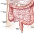 Colostomy closure - reconstructive coloplasty