Microtubules where are located. The structure of microtubules and their functions. The scheme of the structure of microtubule
Polar: At one end, the self-consumption of microtubule occurs, on the other - disassembly. In microtubule cells play the role of structural in many cell processes.
Structure
Microtubules are structures in which 13 protofilaments consisting of α-and β-tubulin heterodimers are laid around the circumference of the hollow cylinder. The outer diameter of the cylinder is about 25 nm, internal - about 15.
One of the ends of the microtubule, called a plus-end, constantly joins the free tubulin. From the opposite end - minus end - tubuline units are cleaved.
β-tubulin
In the formation of microtubule, three phases are isolated:
- Slowd phase, or nucleation. This is the phase of the origin of the microtubule, when the tubulin molecules begin to connect to larger formations. Such a connection takes slower than the addition of tubulin to the already assembled microtubule, so the phase is called slow motion.
- Phase polymerization, or elongation. If the concentration of free tubulin is high, its polymerization occurs faster than the depolymerization on the minus end, due to which the microtubule is extended. As it grows, the concentration of tubulin falls to the critical, and the growth rate slows down until joined the next phase.
- Phase of stable state. Depolymerization balancing polymerization, and microtubule growth stops.
Laboratory studies show that the assembly of microtubules from tubulins occurs only in the presence of Guanosintriphosphate and magnesium ions.
Video on the topic
Dynamic instability
Microtubule are dynamic structures and in the cell are constantly polymerized and depolymerized. Centrosoma, localized near the nucleus, performs in animal cells and many rubbers as the center of the organization of microtubule (COMT): they grow from it to the periphery of the cell. At the same time, microtubules can suddenly stop their growth and shorten back towards the centrosome to complete destruction, and then grow again. When connecting to the microtube, a tubulin molecule, carrier GTF, form a "cap", which provides the growth of microtubule. If the local concentration of tubulin falls, associated with beta tubulin GTF gradually hydrolyzed. If the GTF "caps" is completely hydrolyzed by the GTF, it leads to a rapid decay of microtubule. Thus, the assembly and disassembly of microtubules is associated with the cost of GTF energy.
The dynamic instability of microtubule plays an important physiological role. For example, when dividing the cells of microtubules grow very quickly and contribute to the correct orientation of chromosomes and the formation of mitotic spindle.
Function
Microtubules in the cell are used as "rails" for the transportation of particles. On their surface, membrane bubbles and mitochondria can move. Transportation on microtubules is carried out proteins called motor. These are high molecular weight compounds consisting of two heavy (weighing about 300 kDa) and several lung chains. In severe chains, head and tail domains are isolated. Two head domains are associated with microtubules and are actually engines, and tailings are associated with organelles and other intracellular formations subject to transportation.
Two types of motor proteins are distinguished:
- cytoplasmic dineins;
Organization and dynamics
Microtubules are excessively sensitive to biotic and abiotic environmental factors (cold, lighting, drought, salinization, the effect of herbicides and pesticides, flooding, compression, exposure to the electric field, pressure and strength of gravity), as well as to phytogorms, antimitotic preparations and a number of other biologically active compounds. Microtubules are hollow polar cylindrical filaments with a diameter of more than 24 nm, which are collected from α-and β-tubulin heterodimers, which in the "head-to-tail" position form 13 protofilaments.
In the cells of higher plants there are four types of microtubule constructions:
Proteins associated with microtubule
All components of the cytoskeleton and other organelles are interconnected by a number of specific proteins associated with microtubes ( BAM). In animal cells, the most studied BAM is tau. and BAM2.which stabilize microtubules and attach them to other cellular structures, as well as transport proteins Dienein and Kinesin. The functioning of various groups of vegetable microtubules depends on the presence of izoforms of the BAM from the family BAM65 and regulatory kinases and phosphatas. In particular, the high-screen animal homolog of the BAM65 family is important for microtubes of certain configurations throughout the plant development. The orientation and organization of various populations and types of microtubule constructions are tissue and organosophic.
Building the root of the tale of Thale Arabidopsis Thaliana L. Typically for bipartite plants. The nearest root surface is the epidermal layer whose cells in the mature zone depending on the ability to initiate the development of root hairs are trichoblasts or atrichoblasts. Deeper there are accumulative non-neuroplast cortical layer with numerous interclausers and plasmodesmas and a layer of endodermal cells with capping belts on anticline surfaces. The central cylinder of the root form parenchymal pericyclas, which are capable of rapid division, and elements of xylems and floems. The functional distinction between the root zones is also present: division zones, elongation, ripening, as well as the transition zone on the border of the initiation and elongation zones. Side roots are formed with pericycle, and with trichoblasts of the epidermal layer - root hairs. The root tip is covered with a root case with specific Morphology of Columella Cells.
Cortical microtubules
Acentrosomal cortical microtubules ( KMT.) Important for morphogenesis of plants, regulation of cellular division and elongation. The highly dynamic population of membrane-bound short CMT is quickly reorienting from an interphase transverse position into a longitudinal cell during elongation of the cell. Acentroscomal cortical microtubules have disordered placement of plus ends and detect dynamic instability, and the free minus ends KMT slowly depolymerizes, that is, the KMT is self-organized by the hybrid mechanism of dynamic instability and Tredaming. Enucleation takes place throughout the surface of the plasma membrane. SPR1 protein regulates the dynamics and organization of plus-terminus KMT plants, which affects anisotropic cell growth. Acentrous cortical microtubules are located parallel to cellulose microfibrillers
Microtubules support the structure of the cells, since they are the most durable of the polymers of the cytoskeleton. Microtubules are resistant to compression
The cage occurs dynamic assembly processes and disassembling microtubules, allowing you to quickly reorganize the cytoskeleton
Cells can provide greater or smaller dynamics of the nature of the functioning of microtubules, providing them with large adaptive capabilities (with a more dynamic nature) or increasing their stability (at a more stable character)
In accordance with specific requirements, various cells are characterized by a specific organization of microtubule
Simple experiments in which microtububles are destroyedillustrate the importance of these components of the cytoskeleton. Under the action of substances similar to colchiscin associated together to the tubulin subunits can depolymerize. These substances block the formation of new microtubules, causing an imbalance between constant formation and decay of the components of the cytoskeleton. Microtubule, which were subjected to depolymerization, cannot recover, which soon leads to the loss of all microtubes in the cytoplasm.
Usually, with depolymerization of microtubuleMost cells acquire a shape. Also violated the internal organization of cells. The Golgi complex, which is usually localized next to the kernel in the form of a discrete structure, forms scattered by the cell fragments. EPR, which represents a network that penetrates the entire cytoplasm is collected around the core, since it is associated with a nuclear shell.
When removing substances, depolymerizing microtubulesAll these changes pass. The microtubule network is restored, the cell shape is returned, and the EPR and the Golgi apparatus occupy the previous position. This simple experiment illustrates the functions of microtubules in the organization of the structure and mobility of the cells.
In a cage microtubule functions They are determined by two properties that have contradictory nature: microtubules can act as rigid structural elements, and they are at the same time they can easily collapse. The structure of the structure and relatively large diameter of the microtubules provide their relative rigidity and resistance to compression. In this regard, they resemble a plumbing hose, which can be engaged at a considerable extent, but it does not compress. However, in contrast to the hose, microtubule is characterized by extremely dynamic nature.
The picture made in the fluorescent microscope is shown about 12 cells, in which microtubule and chromosome are visible.One of the mitotic cells in which microtubules are assembled into mitotic spindle, is surrounded by interphase cells.
Reorganization of microtubules, which occurs when the cell entry into mitosis is taken, is deep, but requires only a few minutes.
They are constantly increasing or shortening due to adding or loss subunits. The shortening of microtubules can have particularly significant consequences, since it often significantly reduces their length, up to a complete disappearance. After the microtubule assembly is characterized by dissociation, and often the cell uses other proteins to stabilize them and prevent this process. Although the property of collapse seems strange for structural elements, such instability has a great advantage, allowing microtubule, if necessary, to understand and reorganize over minutes.
An example of this is a complete restructuring microtubule networkswhich occurs at the beginning of mitosis and takes just a few minutes. Another example is the reorganization of microtubule, which occurs in developing oocytes and illustrates the extensive nature of this process. The figure below shows the Xenopus Laevis Frog Oocytes. In diameter, it is about 1 mm in diameter and contains approximately half a million microtubules, the average length of which reaches 600 μm. If all the subunits of these microtubes pull into one continuous line, then it will be 300 m, that is, three football fields. Despite such a large number of microtubules, when the oocyte is stimulated to ripen in the egg, the entire cytoskeleton depolymerizes and is reorganized within 30 minutes.
For some cells dynamic nature microtubule means more than just the ability to quickly transition of one type of cytoskeleton in another. For example, a fibroblast must have the ability to move in the body and at the same time change the direction of movement. In these cells, microtubules are organized in the form of radial radial structures that apply from one point, close to the kernel.
These microtubule There are not long, for only part of the time required for the movement of the cell for a certain distance. Fibroblast can continue to move, even if all microtubes have undergone depolymerization. It is interesting, however, that without microtubes, the cell is not able to change the direction of movement. Apparently, this requires the dynamic nature of microtubules.
Neuron It is very different in form and on behavior from the fibroblast. Neuron is a fixed cell, which is characterized by a small body and protruding from it by the processes (axon and dendrites) spread over long distances. Inside the processes, the microtubule system passes. These microtubule carry a large number of vesicles and other materials to synapses and in the opposite direction. Unlike microtubules present in Fibroblast, neuron processes are stable and play a major role in ensuring the structure of the cell.
 The Mature Xenopus Laevis Oocyte is a large cell in which microtubule is tightly packed.
 In two photos, microtubule are presented close to the edges of the oocyte.
Despite the large number of microtubule present and the fact that their total length reaches a significant amount,
they are able to fully figure out for a few minutes.
For depolymerization There is a slow destruction of processes. Thus, neurons use the features of the structure of microtubules to create stable structural elements.
Although mature neurons Microtubules use to strengthen their structure, growing neurons also use the dynamic properties of microtubules. When neurons begin to grow and form synapses with other neurons, their cellular bodies form subtle increases that become axon and dendrita. At the end of each grow out there is a very active moving area, which is called a growth cone and which can be seen in the figure below. Growth cones apply to long distances, and as they have been promoted behind them. Growth cones contain dynamic microtubules that function in the same way as in moving fibroblasts, and contribute to the movement of growth cones.
In this way, neurons They themselves decide on what moments and in what places the cells of the microtubule should acquire dynamic character, and when they should remain stable. The ability to adjust the dynamic state of microtubules in time and space is the common property of all cells.
Microtubule. Organized in accordance with the individual needs of each cell. The figure below shows two close in the form of cell type: single-celled schizosaccharomyces pombe yeast, and nuclear erythrocytes of some vertebrates, such as birds and amphibians. In the other case, the cells have an oblong form, but they are organized in different microtubule. In S. pombe cells, microtubule bundles are oriented longitudinally and directed to the cell edge, where the components needed for the polar cell growth are accumulated. Microtubule bundles also position the kernel in the center of the cell.
In cells S. POMBE. Microtubule does not perform a protective function from mechanical effects, since they are protected by a cell wall. In red blood cells, a completely different organization of microtubules is found, since they, as in all animal cells, there is no cell wall. These cells have bunches of microtubules combined together with the plasma membrane into the structure located along the periphery of the cell (t. N. Marginal beam).
 The various form of two types of cells requires different organization of their microtubules.
 In the fibroblast of a person, individual microtubules are visible, which begin at a point located near the nucleus, and pass through the cytoplasm.
In neuron, microtubules are packed together in long subtle formations, which come out of the body of the cell.
Microtubule. marginal beam provide the rigidity of the cell membrane; The same function in the red blood cells of mammals have an aquerine and spectrine proteins.
Presented examples Illustrate the general functions of microtubules and generate a lot of questions. How are microtubule capable of so quickly assess and dissociate? How do cells regulate the dynamics of assembly and disassembly microtubules? What determines the organization of microtubules in the cell?
General characteristics of microtubules.The mandatory components of the cytoskeleton include microtubules (Fig. 265), nichly unreasonable structures, 25 nm thick, consisting of protein-tubulins and proteins associated with them. Polymerization tubulines form hollow tubes (microtubule), the length of which can reach several microns, and the longest microtubules are found as part of the axes of sperm tails.
Microtubules are located in the cytoplasm of interphase cells, by one, small loose beams, or in the form of tightened formations in the center of centrioles, basal taurus in cilia and flagella. When cell dividing the cells, most microtubes cells are part of the separation spine.
On the structure of the microtubules are long hollow cylinders with an outer diameter of 25 nm (Fig. 266). The wall of the microtubule consists of polymerized tubulin protein molecules. In the polymerization of the tubulin molecule, 13 longitudinal protofilaments are formed, which twisted into the hollow tube (Fig. 267). The size of the tubulin monomer is about 5 nm equal to the thickness of the microtubule wall, in the cross section of which 13 globular molecules are visible.
The tubulin molecule is a heterodimer consisting of two different subednits, from a-tubulin and b-tubulin, which in the association form the tubulin protein itself, originally polarized. Both subunits of a monomer of tubulin are associated with GTF, but the GTF is not subjected to hydrolysis, unlike GTF on the B-subunit, where, with polymerization, GTF hydrolysis takes place to GDF. In the polymerization of the tubulin molecule, they are combined in such a way that with the B-subunit of one protein associates A-subunit of the next protein, etc. Consequently, individual protofibrils arise as polar threads, and, accordingly, the entire microtubule is also a polar structure having a rapidly growing (+) - the end and slowly growing (-) end (Fig. 268).
With a sufficient concentration of protein, polymerization occurs spontaneously. But with the spontaneous polymerization of tubulins, the hydrolysis of a single GTF molecule associated with B-tubulin occurs. During the increase in the length of the microtubule, the binding of tubulins occurs with a greater speed on the growing (+) - end. But with an insufficient concentration of tubulin, the microtubules can deal with both ends. Disassembling microtubules contributes to a decrease in temperature and the presence of Ca ++ ions.
Microtubules are very dynamic structures that can quickly arise and understand. As part of the isolated microtubules, additional proteins associated with them are found, so-called. Mar-proteins (Mar- Microtubule Accessory Proteins). These proteins, stabilizing microtubule, accelerate the process of polymerization of tubulin (Fig. 269).
The role of cytoplasmic microtubules is reduced to the execution of two functions: skeletal and motor. Skeletal, frame, the role is that the location of microtubules in the cytoplasm stabilizes the shape of the cell; When the microtubule is dissolved, the cells that had a complex shape tend to acquire the shape of the ball. Muscular role of microtubules is not only that they create an ordered, vector, motion system. Microtubules of cytoplasm in association with specific associated motor proteins form ATP-azny complexes capable of moving cell components.
In almost all eukaryotic cells in the hyaloplasm you can see long unreasonable microtubules. In large quantities, they are found in cytoplasmic processes of nerve cells, in melanocyte processes, ameb and other cells that change their shape (Fig. 270). They can be highlighted themselves or you can select their forming proteins: these are the same tubulines with all their properties.
Centers of the organization of microtubule. The growth of the microtubules of the cytoplasm occurs polarly: increasing (+) - the end of the microtubule. The lifetime of microtubules is very short, therefore the formation of new microtubules is constantly. The process of starting the polymerization of tubulins, nucleation, occurs in clearly bounded areas of the cell, in the so-called. The centers of the organization of microtubule (COMT). In the zones of the COMT, there is a bookmark of short microtubules facing its (-) - ends to the COMT. It is believed that in the ZOMT zones (-) - the ends are blocked by special proteins that prevent or limit the depolymerization of tubulins. Therefore, with a sufficient amount of free tubulin, the length of the microtubules derived from the COMT will occur. As a COMT in animal cells, highly cellular centers containing centrioles are involved, which will be mentioned below. In addition, a nuclear zone can serve as a COMT, and during mitosis of the pole spine division.
One of the prescriptions of the microtubules of the cytoplasm is to create an elastic, but at the same time a stable intracellular skeleton necessary to maintain the cell form. The diskheads in the form of erythrocytes amphibians along the periphery of the cell lies the harness of circularly laid microtubules; Bundles of microtubules are characteristic of different cytoplasm growth (the axiopod of the simplest, axons of nervous cells, etc.).
The role of microtubule is to form a frame for maintaining a cellular body, to stabilize and strengthen cellular growth. In addition, microtubules are involved in cell growth processes. So, in plants in the process of stretching the cells, when a significant increase in the volume of cells occurs due to an increase in the central vacuole, large numbers of microtubes appear in the peripheral layers of the cytoplasm. In this case, microtubule, as well as the cell wall growing at this time, as if reinforced, mechanically strengthen the cytoplasm.
Creating an intracellular skeleton, microtubules are factors of the oriented movement of intracellular components, setting the space for directed flows of different substances and to move large structures. Thus, in the case of melanognors (cells containing the melanin's pigment), the pigment granules increase along the microtubule beams.
In the axons of living nerve cells, you can observe the movement of various small vacuoles and granules, which move both from the body of the cell to the nerve end (anterograde transport) and in the opposite direction (retrograde transport).
Proteins responsible for the movement of vacuoles were allocated. One of them is a kinesin, a protein with a molecular weight of about 300 thousand.
There is a whole family of kinesins. Thus, cytosol kinesins are involved in transport on microtubules vesicle, lysosomes and other membrane organelles. Many of the kinesins are associated specifically with their goods. So some participate in the transfer only mitochondria, others - only synaptic bubbles. Kinesins are associated with membranes through membrane protein complexes - kinectins. Kinesins of the separation of division participate in the formation of this structure and in the discrepancy of chromosomes.
For retrograde transport in the Axone, another protein is replied - cytoplasmic dienein (Fig. 275). It consists of two heavy chains - heads interacting with microtubes, several intermediate and light chains that bind to membrane vacuoles. The cytoplasmic dyerine is a motor protein carrying cargo to the minus end of microtubules. Dieneins are also divided into two classes: cytosol - participating in the transfer of vacuoles and chromosomes, and axonney - responsible for the movement of cilia and flagella.
Cytoplasmic dineins and kinesins were found in almost all types of cells of animals and plants.
Thus, in the cytoplasm, the movement is carried out according to the principle of sliding threads, only along the microtubules are moved not the threads, but short molecules - movements associated with moving cellular components. The similarity with the actomyosine complex of this system of intracellular transport is that a double complex is formed (microtubule + moti), which has a high ATP-azna activity.
As can be seen, microtubule form radially divergent polarized fibrils, (+) - the ends of which are directed from the center of the cell to the periphery. The presence of (+) and (-) - directed motor proteins (kinesins and dinaines) creates the possibility for transferring its components as from the periphery to the center (endocytosis vacuole, recycling of the vacuoles of the ER and the Machinery, etc.) and the center Peripherals (vacuoles ER, lysosomes, secretory vacuoles, etc.) (Fig. 276). This polarity of transport is created at the expense of the organization of the microtubule system arising in the centers of their organization, in the cellular center.
The overall characteristic of the microtubule
One of the required components of the eukaryot cytoskeleton are microtubule(Fig. 265). These are nichly unprecedented structures, 25 nm thick, consisting of tubulin proteins and associated proteins. The tubulines of microtubules for polymerization form hollow tubes, from where and their name. The length of them can reach several microns; The longest microtubules are found in the axes of sperm tails.
Microtubules are found in the cytoplasm of interphase cells, where they are arranged by one or small loose bundles, or in the form of delicated microtubes in the center of centrioles, basal taurus and in cilia and flagella. When cell dividing the cells, most microtubes cells are part of the separation spine.
In the morphological terms, the microtubules are long hollow cylinders with an outer diameter of 25 nm (Fig. 266). The wall of the microtubule consists of polymerized tubulin protein molecules. In the polymerization of the tubulin molecule, 13 longitudinal protofilaments are formed, which twisted into the hollow tube (Fig. 267). The size of the tubulin monomer is about 5 nm equal to the thickness of the microtubule wall, in the cross section of which 13 globular molecules are visible.
The tubulin molecule is a heterodimer consisting of two different subednits, from -tubulin and -tubulin, which, with the association, form the tubulin protein itself, originally polarized. Both lesions of the Tubulin monomer are associated with GTF, however, the GTF is not subjected to hydrolysis, in contrast to the GTF on a subunit, where the polymerization occurs, GTF is hydrolysis to GDF. In the polymerization of the tubulin molecule, they are combined in such a way that with a subunit of one protein associates a subunit of the next protein, etc. Consequently, individual protofibrils arise as polar threads, and, accordingly, the entire microtubule is also a polar structure having a rapidly growing (+) - the end and slowly growing (-) end (Fig. 268).
With a sufficient concentration of protein, polymerization occurs spontaneously. But with the spontaneous polymerization of tubulins, the hydrolysis of one GTF molecule associated with -tubulin occurs. During the increase in the length of the microtubule, the binding of tubulins occurs with a greater speed on the growing (+) - end. But with an insufficient concentration of tubulin, the microtubules can deal with both ends. Disassembling microtubules contributes to a decrease in temperature and the presence of Ca ++ ions.
There are a number of substances that affect the polymerization of tubulin. Thus, the alkaloid colchicine contained in a nonsense autumn-free (Colchicum Autumnale) binds to individual tubulin molecules and prevents them from polymerization. This leads to a drop in the concentration of a free tubulin capable of polymerization, which causes a quick disassembly of cytoplasmic microtubules and microtubes of the separation of division. The same action has a coarse and nocodoxol, when laundering which microtubules are completely restored.
A stabilizing action on microtubule has a taxol, which contributes to the polymerization of tubulin even at its low concentrations.
All this shows that microtubules are very dynamic structures that can quickly arise and understand.
As part of the isolated microtubules, additional proteins associated with them are found, so-called. Mar-proteins (Mar- Microtubule Accessory Proteins). These proteins, stabilizing microtubule, accelerate the process of polymerization of tubulin (Fig. 269).
Recently, the process of assembling and disassembling microtubules began to observe in living cells. After introducing into the cell labeled with fluorochromes of antibodies to a tubuline and using electronic signal amplification systems in a light microscope, it can be seen that in a lively cell microtubules grow, stuffed, disappear, i.e. constantly being in dynamic instability. It turned out that the average half-life of cytoplasmic microtubules is only 5 minutes. So in 15 minutes about 80% of the entire population of microtubules is updated. At the same time, individual microtubules may be slow (4-7 μm / min) to lengthen, and then quite quickly (14-17 μm / min) to shorten. In the living cells of the microtubule in the composition of the separation of division have a lifetime of about 15-20 seconds. It is believed that the dynamic instability of cytoplasmic microtubules is associated with the delay in hydrolysis GTF, this leads to the fact that on (+) - the end of the microtubule is formed by a zone containing nonhydrolyzed nucleotides ("GTF cap"). In this zone, the tubulin molecule is associated with great affinity for each other, and, therefore, the growth rate of microtubule increases. On the contrary, with the loss of this plot, microtubule begin to shorten.
However, 10-20% of microtubules remain relatively stable enough long time (up to several hours). Such stabilization is observed in a large degree in differentiated cells. The stabilization of microtubules is associated or with a modification of tubulins or with their binding with additional (ID) proteins of microtubule and with other cellular components.
Lisin acetylation in the composition of tubulins significantly increases the stability of microtubules. Another example of modification of tubulins may be the removal of terminal tyrosine, which is also characteristic of stable microtubules. These modifications are reversible.
Microtubule themselves are not capable of reduction, however, they are the required components of many moving cellular structures, such as cilia and flagellas, such as spindle cells during mitosis, as microtubules of cytoplasm, which are mandatory for a number of intracellular transports, such as exocytosis, mitochondrial movement, etc. .
In general, the role of cytoplasmic microtubules can be reduced to two functions: skeletal and motor. Skeletal, frame, the role is that the location of microtubules in the cytoplasm stabilizes the shape of the cell; When the microtubule is dissolved, the cells that had a complex shape tend to acquire the shape of the ball. Muscular role of microtubules is not only that they create an ordered, vector, motion system. Microtubules of cytoplasm in association with specific associated motor proteins form ATP-azny complexes capable of moving cell components.
In almost all eukaryotic cells in the hyaloplasm you can see long unreasonable microtubules. In large quantities, they are found in cytoplasmic processes of nerve cells, in melanocyte processes, ameb and other cells that change their shape (Fig. 270). They can be highlighted themselves or you can select their forming proteins: these are the same tubulines with all their properties.
Centers of the organization of microtubule.
The growth of the microtubules of the cytoplasm occurs polarly: increasing (+) - the end of the microtubule. Since the lifetime of microtubule is very short, then the formation of new microtubules should constantly occur. The process of starting the polymerization of tubulins, nucleation, occurs in clearly bounded areas of the cell, in the so-called. centers of organization microtubule (COMT). In the zones of the COMT, there is a bookmark of short microtubules facing its (-) - ends to the COMT. It is believed that in the ZOMT zones (-) - the ends are blocked by special proteins that prevent or limit the depolymerization of tubulins. Therefore, with a sufficient amount of free tubulin, the length of the microtubules derived from the COMT will occur. Mainly cell centers containing centrioles are involved in the cells of animals, which will be said later. In addition, a nuclear zone can serve as a COMT, and during mitosis of the pole spine division.
The presence of centers of the organization of microtubules is proved by direct experiments. So, if in the living cells to completely depolymerize microtubule or using a rolleride or by cooling the cells, then after removing the effects, the first signs of the appearance of microtubes will appear in the form of radially diverging rays that are separated from one place (citner). Usually in cells of animal origin, the citner occurs in the zone of the cellular center. After such primary nucleation, the microtubule begin to grow from the COMT and fill the entire cytoplasm. Consequently, the growing peripheral ends of microtubules will always (+) - ends, and (-) - the ends will be located in the zone of the COMT (Fig. 271, 272).
Cytoplasmic microtubules arise and diverge from one cellular center with which many people lose touch can quickly understand, or, on the contrary, can stabilize with association with additional proteins.
One of the functional prescriptions of the microtubules of the cytoplasm is to create an elastic, but at the same time a stable intracellular skeleton required to maintain the shape of the cell. It was found that the discosites in the form of the erythrocytes of amphibians along the periphery of the cell lies the harness of circularly laid microtubules; Bundles of microtubules are characteristic of different cytoplasm growth (the axiopod of the simplest, axons of nervous cells, etc.).
The action of a colchicine causing the depolymerization of tubulins strongly changes the shape of the cell. So, if the grip and flat cage in the culture of fibroblasts to treat a colchicine, then it loses polarity. Other cells behave in exactly the same way: colchicine stops the growth of lens cells, nerve cell processes, muscle tubes, etc. Since it does not disappear the elementary forms of inherent motion cells, such as pinocytosis, adulting the movements of membranes, the formation of small pseudopodies, then the role of microtubule is to form a frame for maintaining a cellular body, to stabilize and strengthen cellular growth. In addition, microtubules are involved in cell growth processes. So, in plants in the process of stretching the cells, when a significant increase in the volume of cells occurs due to an increase in the central vacuole, large numbers of microtubes appear in the peripheral layers of the cytoplasm. In this case, microtubule, as well as the cell wall growing at this time, as if reinforced, mechanically strengthen the cytoplasm.
Creating such an intracellular skeleton, microtubule can be factors of the oriented motion of intracellular components, set as its location of space for directed flows of different substances and to move large structures. Thus, in the case of melanognors (cells containing the melanin's pigment), the pigment granules increase along the microtubule beams. The destruction of microtubules with colchicine leads to a violation of the transport of substances in the axons of nerve cells, to the termination of exocytosis and the blockade of secretion. In the destruction of the microtubules of the cytoplasm, fragmentation occurs and running around the cytoplasm of the Golgi apparatus, the destruction of mitochondrial reticulum.
For a long time it was believed that the participation of microtubule in the movement of cytoplasmic components is only that they create a system of ordered movement. Sometimes in popular literature, cytoplasmic microtubules are compared with railway rails, without which the movement of trains is not possible, but which they themselves do nothing in themselves. At one time it was assumed that the engine, locomotive, can be a system of actin filaments, but it turned out that the mechanism of intracellular movement of various membrane and non-smuggled components is associated with a group of other proteins.
Progress was achieved when studying the so-called. Axonial transport in giant neurons of squid. Axons, nerve cell processes may have a large length and filled with a large number of microtubule and neurofilaments. In the axons of living nerve cells, you can observe the movement of various small vacuoles and granules, which move both from the body of the cell to the nerve end (anterograde transport) and in the opposite direction (retrograde transport). If the axon is dragging out the thin ligature, then such transport will lead to a cluster of small vacuoles on both sides of the tank. Vacuols moving Anteorograde contain various mediators, in the same direction can move and mitochondria. Retrogradi moves vacuoles formed as a result of endocytosis when recycling membrane sites. These movements occur with relatively high speed: from the neuron body - 400 mm per day, in the direction to Neurone -200-300 mm per day (Fig. 273).
It turned out that from the segment of a giant axon of a squid can be allocated ashlasm, the contents of the axon. In a drop of isolated axoplasm, the movement of small vacuoles and granules continues. With the help of a video concessive device, it can be seen that the movement of small bubbles occurs along thin pitchal structures along the microtubule. Of these drugs, proteins responsible for the movement of vacuoles were allocated. One of them kinesin, protein with molecular weight about 300 thousand. It consists of two similar heavy polypeptide chains and several lungs. Each heavy chain forms a globular head, which, with association with a microtubule, has ATP-azna activity, while light chains are binding to the membrane of bubbles or other particles (Fig. 274). In the hydrolysis of ATP, the conformation of the kinesin molecule changes and the particle moves in the direction to (+) - the end of the microtubule. It turned out to be possible to glue, immobilize kinesin molecules on the surface of the glass; If add free microtubule in the presence of ATF to this drug, the latter begin to move. On the contrary, you can immobilize microtubules, but add membrane bubbles associated with kinesine - bubbles begin to move along microtubules.
There is a whole family of kinesins with similar motor heads, but characterized by tail domains. Thus, cytosol kinesins are involved in transport on microtubules vesicle, lysosomes and other membrane organelles. Many of the kinesins are associated specifically with their goods. So some participate in the transfer only mitochondria, others - only synaptic bubbles. Kinesins are associated with membranes through membrane protein complexes - kinectins. Kinesins of the separation of division participate in the formation of this structure and in the discrepancy of chromosomes.
For retrograde transport in Axone answers another protein - cytoplasmic dienein(Fig. 275).
It consists of two heavy chains - heads interacting with microtubes, several intermediate and light chains that bind to membrane vacuoles. The cytoplasmic dyerine is a motor protein carrying cargo to the minus end of microtubules. Dieneins are also divided into two classes: cytosol - participating in the transfer of vacuoles and chromosomes, and axonney - responsible for the movement of cilia and flagella.
Cytoplasmic dineins and kinesins were found in almost all types of cells of animals and plants.
Thus, in the cytoplasm, the movement is carried out according to the principle of sliding threads, only along the microtubules are moved not the threads, but short molecules - movements associated with moving cellular components. The similarity with the actomyosine complex of this system of intracellular transport is that a double complex is formed (microtubule + moti), which has a high ATP-azna activity.
As we can see, microtubule form in the cell radially divergent polarized fibrils, (+) - the ends of which are directed from the center of the cell to the periphery. The presence of (+) and (-) - directed motor proteins (kinesins and dinaines) creates the possibility for transferring its components as from the periphery to the center (endocytosis vacuole, recycling of the vacuoles of the ER and the Machinery, etc.) and the center Peripherals (vacuoles ER, lysosomes, secretory vacuoles, etc.) (Fig. 276). This polarity of transport is created at the expense of the organization of the microtubule system arising in the centers of their organization, in the cellular center.
The structure and functions of microtubules.
One of the mandatory components of the cytoplasm of the plant cell are microtubules. In the morphological terms, the microtubules are long hollow cylinders with an external diameter of 25 nm. The wall of the microtubule consists of polymerized tubulin protein molecules. In the polymerization of the tubulin molecule, 13 longitudinal protofilaments are formed, which twisted into the hollow tube. The exchange of a tubulin monomer is about 5 nm equal to the thickness of the microtubule wall, in the cross section of which 13 globular molecules are visible.
The microtubule is a polar structure having a rapidly growing plus end and slowly growing minus-end.
Microtubules are very dynamic structures that can quickly arise and understand. When using electronic signal amplification systems in the light microscope, it can be seen that in a lively cell microtubule grow, stuffed, disappear, i.e. constantly being in dynamic instability. It turned out that the average half-life of cytoplasmic microtubules is only 5 minutes. So in 15 minutes about 80% of the entire population of microtubules is updated. As part of the spinner of the microtubor division, there are about 15-20 s. However, 10-20% of microtubules remain relatively stable enough long time (up to several hours).
Microtubule themselves are not able to reduce, but they are the required components of many moving cellular structures, such as spindle cells during mitosis as microtubule cytoplasm, which are mandatory for a number of intracellular transports, such as exocytosis, mitochondrial movement, etc.
In general, the role of cytoplasmic microtubules can be reduced to two functions: skeletal and motor. Skeletal, frame, the role is that the location of microtubules in the cytoplasm stabilizes the shape of the cell. Motor role microtubule is not only that they create an ordered, vector motion system. Microtubules of cytoplasm A association with specific associated motor proteins form atphase complexes capable of moving cell components. In addition, microtubules are involved in cell growth processes. In plants, in the process of stretching cells, when due to an increase in the central vacuole, a significant increase in the volume of cells occurs, large quantities of microtubes appear in the peripheral layers of the cytoplasm. In this case, microtubules, as well as the growing cells at this time
the wall, as if reinforced, mechanically strengthen the cytoplasm.
Chemical composition of microtubule
Microtubule consist of protein-tubulins and associated proteins. The tubulin molecule is a heterodimer consisting of two different subenitages: from
andWith a sufficient concentration of protein, polymerization occurs spontaneously. With spontaneous polymerization of tubulins, the hydrolysis of a single GTF molecule associated with
- Tubulin. During the increase in the length of the microtubule, the binding of tubulins is at a greater speed on the growing plus end. But with an insufficient concentration of tubulin, the microtubules can deal with both ends. Disassembly of microtubes contribute to the temperature and the presence of ions Ca 2.There are a number of substances that affect the polymerization of tubulin. Thus, the alkaloid colchicine is binding to individual tubulin molecules and the ripped possesses their polymerization. This leads to a drop in the concentration of a free tubulin capable of polymerization, which causes a quick disassembly of cytoplasmic microtubules and microtubes of the separation of division. In the same action, the coarse and nocodoxol possess, during the laundering of which the microtubule was completely restored.
A stabilizing action on microtubule has a taxol, which contributes to the polymerization of tubulin even at its low concentrations.
Also as part of microtubules, additional proteins associated with them are found, the so-called mar-proteins. These proteins stabilizing microtubule, accelerate the process of the polymerization of tubulin.
The structure and functions of microfilaments
Microfilaments are very subtle and long filamentine protein structures found in the entire cytoplasm. Under the plasma membrane, microfilaments form a solid plexus, forming a cytoscile. This whole structure is very labil. Under the influence of various impacts (the calcium concentration is of great importance) microfilaments disintegrate into separate fragments and are collected again. Since microfilaments are reduced elements of the cytoskeleton, they are involved in changing the cell shape, in intracellular displacements of the organelle, discrepancies with chromosomes during cell division. In addition, microfilants perform the functions:
Responsible for moving: chloroplasts that can change their position depending on lighting;
Cell cores;
Bubbles;
Participate: in phagocytosis (but not in pin or exocytosis); in the formation of tugs with cellular division (there is a ring from microfilament beams, slimming cells); In the movement of chromatids and chromosomes when dividing the kernel.
Intracellular movement occurs when the interaction of microfilaments from actin (actin yarn) with myosin.
Chemical composition of microfilaments
Microfilaments included in the main actin protein. But besides him, Miosin, Aktinin and others come.
Aktin is a globular protein, it is 5-15% of the entire cell protein and is an essential protein of eukaryotic cells. The globular actin (gamma actin) is polymerized into actin filaments (F-actin), consisting of two spirals twisted near each other (diameter - about 6 nm, length - several microns). Aktin forms a three-dimensional network from a large number of threads or a bundle of at least 20 yarns. In the cell there is a reversible balance: Gamma Aktin - F-Aktin - F-actin beams.
Myozin in eukaryotic cells is contained in less (0.3-1.5% cell protein) than actin. The filamentine molecule of myosin (molecular weight of more than 450,000, length 150 nm) consists of two large and several small subunits forming a long double helix. One end of this helix carries two heads. The end with the head catalyzes the splitting of ATP (myozic atpasis) and can specifically bind to actin. Aktin activates the ATPase. When splitting ATP, the energy required for intracellular movements is released.
Conclusion
The cell wall of plants performs a number of important functions. Surrounding the plant cell on all sides, it serves as a link between it and adjacent cells. Connecting with each other with thin threads of the cytoplasm - plasmodesmas, through which the substances of the cells are moved from the cell.
Due to the fact that the primary shell of elastic, the cell in this period is growing intensively. After the cessation of growth, a secondary shell is formed, which includes lignin and a number of other substances - a cell strength, rigidity. These properties are especially important for land plants: first, it is a durable "skeleton", secondly, protection against excessive water loss. The cell shell is transparent, so the sun's rays easily penetrate the cell to chloroplasts.
The cytoskeleton is protein, unreasonable polymers involved in the process of moving cell components, as well as perform a frame skeletal role. Also, these components are involved in the process of cell division, forming the filament of the separation of division.
From the above it is clear that these cell components play an important
Bibliography
1. Andreeva T.F. Maevskaya S.N. Voevodskaya S.Yu. "Plant Physiology"
2. Heath TC Dobrykh E.V. "Plant Physiology" 1993
3. Frei-Vismeng A. Mületal K. "Ultrastructure vegetable
cells »1968
4. Chentsov Yu.S. "Introduction to cellular biology", M. Academkniga,
5. Yakushkin N.I. Bakhtenko E.Yu. "Plant Physiology", M. Vlados
6. www.ido.tsu.ru.
7. http://www.medkurs.ru/lecture1k/med_bioLogy/Qm31/2499.htm.
8. http://school.iot.ru/predmety/biologiya.doc.
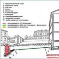 What is a hot water supply of an apartment building
 Water supply of an apartment building
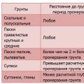 Calculation of the Load for the foundation Installed Electrical Instruments