Fast and slow variables. What periods are distinguished in the development of germ cells? Tell us about the maturation period (meiosis). Reduction of chromosomes in meiosis occurs in
Meiosis(from the Greek. meiosis - reduction) - the process of division of the cell nucleus with the formation of four daughter nuclei, each of which contains half the number of chromosomes than the original nucleus. Meiosis is a reduction division: the number of chromosomes in a cell decreases from diploid (2 n) to haploid (n). Meiosis accompanies the formation of gametes in animals and the formation of spores in plants. As a result of meiosis, haploid nuclei are obtained, when they merge during fertilization, the diploid set of chromosomes is restored
Meiosis (outline). As a result of meiosis, four gametes arise with differing haploid sets of chromosomes (Harnden, 1965).
Meiosis involves two consecutive divisions... In each meiotic division, four stages are distinguished: prophase, metaphase, anaphase and telophase.
The first meiotic division is called reduction. As a result, from one cell with a diploid set of chromosomes, two with a haploid set are formed.
Prophase I, the prophase of the first meiotic division, is the longest. It is conventionally divided into five stages: leptotene, zygotene, pachytene, diplotene and diakinesis.
The first stage, leptotene, is characterized by an enlargement of the nucleus. A diploid set of chromosomes is visible in the nucleus. Chromosomes are long, thin filaments. Each chromosome consists of two chromatids. Chromatides have chromomeric
structure. Spiralization of chromosomes begins.
During the second stage of prophase of the 1st meiotic division - zygotene - conjugation of homologous chromosomes occurs. Homologous chromosomes are called chromosomes that have the same shape and size: one of them is received from the mother, the other from the father. Homologous chromosomes are attracted and attached to each other along their entire length. The centromere of one of the paired chromosomes is exactly adjacent to the centromere of the other, and each chromatid is adjacent to the homologous chromatid
The third stage, pachytene, is the thick filament stage. The conjugating chromosomes are closely adjacent to each other. Such doubled chromosomes are called bivalents. Each bivalent consists of a four (tetrad) chromatids. The number of bivalents is equal to the haploid set of chromosomes. Further spiralization takes place. Close contact between chromatids makes it possible to exchange identical regions in homologous chromosomes. This phenomenon is called crossing over.
The fourth stage - diplotene - is characterized by the appearance of repulsive forces. The chromosomes that make up the bivalents begin to drift apart. The divergence begins in the area of centromeres. Chromosomes are connected to each other at several points. These points are called chiasma (from the Greek chiasma -cross), that is, places where crossing over will occur. In each chiasm, there is an exchange of chromatid regions. Chromosomes spiralize and shorten.
The fifth stage - diakinesis - is characterized by maximum spiralization, shortening and thickening of chromosomes. The repulsion of chromosomes continues, but they remain connected in bivalents at their ends. The nucleolus and nuclear envelope dissolve. The centrioles diverge towards the poles.
Thus, in the prophase of the 1st meiotic division, three main processes take place:
1) conjugation of homologous chromosomes;
2) the formation of bivalents of chromosomes or tetrads of chromatids;
3) crossing over.
Metaphase I. In the metaphase of the first meiotic division, bivalents of chromosomes are located along the equator of the cell, forming a metaphase plate. Fission spindle threads are attached to them.
Anaphase I. In the anaphase of the first meiotic division, chromosomes, not chromatids, diverge to the poles of the cell. Only one of a pair of homologous chromosomes gets into daughter cells.
Telophase I. In the telophase of the first meiotic division, the number of chromosomes in each cell becomes haploid. Chromosomes are composed of two chromatids. Due to crossing over during the formation of chiasmata, chromatids are not genetically homogeneous. For a short time, the nuclear envelope, chromosomes
despiralize, the nucleus becomes interphase. Then, the division of the cytoplasm begins in the animal cell, and the formation of the cell wall in the plant cell. Many plants do not have telophase I, the cell wall is not formed, there is no interphase II, the cells immediately move from anaphase I to prophase II.
Interphase II. Only animal cells have this stage. During the interphase between the first and second division in the S period, there is no molecule reduplication
The second meiotic division is called equational. It is similar to mitosis. From chromosomes that have two chromatids, chromosomes are formed, consisting of one chromatid.
Prophase II. In the prophase of the second meiotic division, the chromosomes thicken and shorten. The nucleolus and nuclear envelope are destroyed. A fission spindle is formed.
Metaphase II. In the metaphase of the second meiotic division, the chromosomes line up along the equator. The threads of the achromatin spindle go to the poles. A metaphase plate is formed.
Anaphase II. In the anaphase of the second meiotic division, the centromeres divide and pull the separated chromatids, called chromosomes, to the opposite poles.
Telophase II, In the telophase of the second meiotic division, the chromosomes are despiralized and become invisible. The spindle threads disappear. A nuclear envelope is formed around the nuclei. The nuclei contain a haploid set of chromosomes. The division of the cytoplasm and the formation of a cell wall in plants occurs. From one original cell, four haploid cells are formed.
THE IMPORTANCE OF MEIOSIS
1. Maintaining the constancy of the number of chromosomes. If there was no reduction in the number of chromosomes during gametogenesis, and the sex cells had a haploid set of chromosomes, then their number would increase from generation to generation.
2. During meiosis, a large number of new combinations of non-homologous chromosomes are formed.
3. In the process of crossing over, recombinations of the genetic
material.
Almost all chromosomes that fall into gametes contain regions originating from both the paternal and maternal chromosomes. This achieves a high degree of recombination of the hereditary material. This is one of the reasons for the variability of organisms, which provides material for selection.
Reduction of [number of] chromosomes gametic reduction- reduction of gametes, reduction of [number] chromosomes.
Reducing the number of chromosomes by half against the somatic set; R.g.- an integral part of reduction division (meiosis).
(Source: "The English-Russian Explanatory Dictionary of Genetic Terms." Arefiev VA, Lisovenko LA, Moscow: VNIRO Publishing House, 1995)
See what "chromosome [number] reduction" is in other dictionaries:
Reduction (syn. Haplosis is outdated.) In genetics, the reduction of the somatic number of chromosomes by half; in animals, as a rule, occurs during the formation of germ cells. Selective reduction (syn. Division of maturation selective) P., at which ... ... Wikipedia
gamete reduction- reduction [number] of chromosomes Reducing the number of chromosomes by half against the somatic set; R.g. an integral part of reduction division (meiosis). [Arefiev V.A., Lisovenko L.A. English Russian Explanatory Dictionary of Genetic Terms 1995 407s.] ... ... Technical translator's guide
Gamete reduction. See reduction [number] of chromosomes. (Source: "The English Russian Explanatory Dictionary of Genetic Terms." Arefiev VA, Lisovenko LA, Moscow: VNIRO Publishing House, 1995) ... Molecular biology and genetics. Explanatory dictionary.
I Reduction (lat. Reductio retraction, return, restoration) in biology, reduction in size, simplification of the structure or complete loss of an organ, tissue or cell in the course of historical development (phylogenesis). II Reduction in cytology regeneration ... Medical encyclopedia
REDUCTION- 1. Decrease of organs or tissues (before their disappearance) and often their loss of function in the process of ontogeny or phylogenesis. 2. Decrease in the number of chromosomes in cells as a result of meiosis ... Dictionary of botanical terms
gametic reduction- ANIMAL EMBRYOLOGY HAMETIC REDUCTION - a halving of the number of chromosomes that occurs during meiosis, during the formation of germ cells - gametes ... General Embryology: Glossary of Terminology
- (from the Greek méiosis reduction) reduction division, division of maturation, a method of cell division, as a result of which there is a decrease (reduction) in the number of chromosomes by half and one diploid cell (containing two sets of chromosomes) ... ... Great Soviet Encyclopedia
- (from the Greek meiosis decrease), division of maturation, a special way of cell division, as a result of which there is a reduction (decrease) in the number of chromosomes and the transition of cells from a diploid state to a haploid one; main link of gametogenesis. M opened V. ... ... Biological encyclopedic dictionary
- (from the Greek meiosis reduction) or reduction cell division division of the nucleus of a eukaryotic cell with a decrease in the number of chromosomes by half. It occurs in two stages (reduction and equational stages of meiosis). Meiosis should not be confused with ... ... Wikipedia
An elementary unit of the living. The cell is separated from other cells or from the external environment by a special membrane and has a nucleus or its equivalent, in which the bulk of the chemical information that controls heredity is concentrated. Studying ... ... Collier's Encyclopedia
Meiosis is a method of division of somatic cells (precursors of germ cells), as a result of which there is a decrease (reduction) in the number of chromosomes and the formation of germ cells with a haploid set of chromosomes.
Phases of meiosis:
1 meiotic division (reduction):
It leads to the formation of diploid cells ( 2n4c) haploid cells ( n2c).
Prophase I of meiosis includes several stages:
· Leptotene- the earliest stage in which the spiralization of chromosomes begins, and they become visible under the microscope as long and thin threads;
· Zygotena- the stage characterized by the onset of conjugation of homologous chromosomes, which are combined into a bivalent;
· Paquitena- the stage in which, against the background of the ongoing spiralization of chromosomes and their shortening, crossing over occurs between homologous chromosomes - crossing with the exchange of the corresponding sections;
· Diplotena- a stage characterized by the emergence of repulsive forces between homologous chromosomes, which begin to separate from each other primarily in the centromere region, but remain connected in the areas of the past crossing over - chiasmata;
· Diakinesis- the final stage of prophase I of meiosis, in which homologous chromosomes are held together only at separate points of the chiasm. Bivalents take on a bizarre shape of rings, crosses, eights, etc. ( 2n4c)
Metaphase I of meiosis: assurance of the formation of the fission spindle. Threads connected by centromeres of homologous chromosomes, heading to different poles, establish bivalents in the plane of the fission spindle equator. ( 2n4c)
Anaphase I of meiosis: bivalents are directed to different poles of the fission spindle. In this case, a haploid set of chromosomes, consisting of two chromatids, departs to each pole. ( 2n4c)
Telophase I of meiosis: at the poles of the spindle, a single, haploid set of chromosomes is collected, each of which contains twice the amount of DNA. ( n2c)
Interkinesis: short interval between two meiotic divisions. It differs from interphase in that there is no DNA replication, chromosome doubling and centriole doubling: these processes occurred in the premeiotic interphase and, partially, in prophase I.
Second meiotic (equational) division:
Prophase II of meiosis: dismantling of nuclear membranes, divergence of centrioles to different poles of the cell, formation of fission spindle filaments. ( n2c)
Metaphase II of meiosis: Alignment of dichromatid chromosomes in the equatorial plane of the cell (metaphase plate), attachment of the spindle filaments with one end to the centrioles, the other to the centromeres of the chromosomes. ( n2c)
Anaphase II of meiosis: Division of two-chromatid chromosomes into chromatids and the divergence of these sister chromatids to opposite poles of the cell (in this case, chromatids become independent single-chromatid chromosomes), recombination of chromosomes. ( 2n2s)
Telophase II of meiosis: Decondensation of chromosomes, the formation of nuclear membranes around each group of chromosomes, the disintegration of the fission spindle filaments, the appearance of the nucleolus, division of the cytoplasm (cytotomy) with the formation of two, and as a result of both meiotic divisions - four haploid cells. ( nc)
Recombination is the process of exchanging genetic material by breaking and combining different molecules. In eukaryotes, it usually occurs during crossing over during meiosis, in particular, during the formation of sperm and eggs.
Reduction is a process that takes place during the maturation of male and female genital elements and is reduced to the fact that the amount of dye elements (chromatin or nuclein) in the nucleus of the reproductive cell is halved.
Meiosis is a special type of cell division resulting in the formation of gametes - sex cells with a haploid set of chromosomes. It consists of two divisions - reduction and equational. In each division of meiosis, as in mitosis, prophase, metaphase, anaphase and telophase are distinguished. Replication of chromosomes occurs during the period S-interphase, preceding meiosis I. At this stage, dividing cells are not yet determined to meiosis. Prophase I is divided into several stages: leptotene, zygotene, pachytene, diplotene, diakinesis. Leptotene(stage of thin filaments), thin twisted filaments of chromosomes appear. Zygotena- there is a conjugation of sections of homologous chromosomes, a synaptonemal complex is formed, which is part of the bivalent. In places where chromatids cross, breaks and exchanges of their sections occur - crossing-over. Paquitena(thick filament stage) is characterized by a haploid number of bivalents. At this stage, the chromomeric pattern of chromosomes is well distinguishable. V diplotene the structure of bivalents and the four chromatids that make up each of them are most clearly visible. At this stage, repulsion of homologues begins and chiasmata become distinguishable. In the diplotene, a greater spiralization of chromosomes is noticeable than at the pachytene stage. V diakinesis spiralization increases, the number of chiasmata decreases, bivalents are located along the periphery of the nucleus. Metaphase I... The nuclear membrane collapses and prophase is replaced by metaphase. The nucleoli disappear. Bivalents are located in the equatorial plane of the cell, forming a metaphase plate. At the same time, the chromosomes are strongly spiralized, thickened and shortened. The spiralization of chromosomes continues until anaphase I, when the chromosomes are maximally spiralized. V anaphase I chromosomes diverge to opposite poles. The paternal and maternal centromeres of each bivalent diverge to opposite poles. Reduction of centromeres occurs. Telophase I characterized by the formation of a nuclear membrane and the restoration of the structure of the nucleus. After a short interkinesis (chromosomes do not double), a second division of meiosis is observed. V prophase II the chromosomes become well distinguishable. Metaphase II- chromosomes are aligned along the equator, they have a clearly pronounced double structure and a large degree of spiralization. V anaphase II the divergence of the doubled centromeres occurs, as a result of which the daughter chromatids diverge to different poles. V telophase II 4 haploid nuclei are formed. The biological significance of meiosis. Meiosis is a method of cell division that underlies the reduction of the number of chromosomes: 2n → n. Weismann was the first to note that the reduction of the number of chromosomes in meiosis and subsequent fertilization underlie the maintenance of the constancy of the number of chromosomes of a species from generation to generation. Meiosis also provides combinative variability (meaning for evolution). Since the chromosomes of different bivalents diverge in anaphase 1 independently of each other, this leads to the recombination of the parental chromosome sets.
Differences between mitosis and meiosis. In the prophase of mitosis, the compaction of chromosomes occurs, meiosis is also the conjugation of homologous chromosomes - the formation of bivalents, recombination. In the metaphase of mitosis, chromosomes are located in the equatorial plane, meiosubivalents. Anaphase of mitosis is the divergence of sister chromatids to the poles; meiosis is an independent divergence of homologous chromosomes to the poles of different bivalents. Telophase mitosis - the formation of two identical diploid nuclei in the cell. Meiosis - 4 haploid cells are formed.
B. 9. 25. General characteristics of bryophytes, their life cycle. Department system. Origin of bryophytes. Mossy - an extensive group of higher plants, very different in external structure. All over the world there are about 25 thousand species. Among the higher plants in terms of the number of species, they occupy the second place after flowering. Mossy are a very ancient group in the plant kingdom. Almost all of them are perennials. Usually mosses are undersized: their height ranges from a few millimeters to 20 cm. They always grow in places of high humidity. Among bryophytes, two large classes are distinguished - Liverworts and Leafy mosses.
In liverworts, the body is represented by a branched green flat thallus. In leafy mosses, the stems and small green leaves are clearly visible, that is, there are shoots. Both have rhizoids that absorb water from the soil and anchor the plants. All bryophytes are characterized by a significant simplicity of the internal structure. In their body there are basic and photosynthetic tissues, but there are no conductive, mechanical, storage and integumentary tissues. It is characterized by the predominance of the haploid gametophyte over the diploid sporophyte in the cycle. Individual life of bryophytes with spore germination. When the exine spore swells, it bursts, and the intine, together with the contents of the spore, stretches out in the form of a papilla, which dividing gives rise to either a single-row filament or a single-layer plate carrying the rhizoid. This is the initial stage of the gametophyte - the stage of the protonema. It is divided into a green assimilating part - chloronema, and a colorless underground part - rhizoderm. The epidermis of thallus and leafy bryophytes is devoid of cuticles and typical stomata; there are no sieve tubes and tracheids in the conducting system. They are characterized by absorption not so much physiologically as physically: due to capillarity, hygroscopicity, swelling. The origin is attributed to the end of the Devonian, the beginning of the Carboniferous, and is divided into 3 classes - Liverworts, Anthocerotes and Leafy mosses. The classification is based on the structure of the body, gametophytes, structural features of the rhizoids, the structure and nature of the opening of the capsules and the geographical location. Thallus of marshantia is flat, branched in the form of lobes, from above the thallus is covered with a single-layer epidermis with stomata. Photosynthetic tissue is divided into air chambers by partitions. Thallus adheres tightly to the substrate with the help of rhizoids. On male gametophytes, the antheridia are located on the upper side of the support, and on female gametophytes, the archegonia are located on the lower side of the support. After fertilization, a sporophyte develops from the formed zygote in the form of a capsule on a short stalk. Before the maturation of the spores in the capsule, a reduction division occurs, the spores in the sporangia are loosened with specialized threads - elasters and thrown out. Germinating spores give rise to a haploid gametophyte in the form of a lamellar protonema.
26. The interaction of neurons in the nerve centers. Interaction between the processes of excitation and inhibition. Reflex and reflex arc concept. Mono- and polysynaptic reflexes. The property of the nervous tissue to transmit excitation by conduction. Excitation is carried out along the nerve fibers in isolation and does not pass from one fiber to another, which is impeded by the sheaths covering the nerve fibers. The excitation is based on a change in the concentration of ions on both sides of the nerve cell membrane. The activity of the nervous system is of a reflex nature. The response to stimulation by the nervous system is called a reflex. The path through which nervous excitement is perceived and transmitted to the working organ is called a reflex arc. It consists of 5 sections: 1) a receptor that perceives irritation, 2) a sensory (centripetal) nerve that transmits excitation to the center, 3) a nerve center, where excitation switches from sensory neurons to motor ones, 4) a motor (centrifugal) nerve that carries excitation from the central nervous system to the working body, 5) the working body that responds to the received irritation. The process of inhibition is the opposite of arousal: it stops activity, weakens or prevents its occurrence. Excitation in some centers of the nervous system is accompanied by inhibition in others: nerve impulses entering the central nervous system can delay certain reflexes. Both processes - excitation and inhibition - are interconnected, which ensures the coordinated activity of organs and the whole organism as a whole. For example, while walking, the contraction of the flexor and extensor muscles alternates: when the flexion center is excited, the impulses follow to the flexor muscles, at the same time the extension center is inhibited and does not send impulses to the extensor centers, as a result of which the latter relax, and vice versa. To perform their functions - to perceive information, process it and transmit a motor impulse to the executive organ - the processes of nerve cells form special connections with neurons of other cells - synapses. When a signal arrives at the end of the axon, a chemical substance is released there, which causes excitation or inhibition in a neighboring cell. Such substances are called mediators, for example, acetylcholine, norepinephrine, etc.
27. Morphology and functions of cellular forms of loose connective tissue. Reticulin, elastic and collagen fibers. Their microscopic structure, physical properties, chemical composition. Connective tissue, in a cut of cells there are still relatively many, and the intercellular substance is not so rich in fibers called. loose connective tissue. It is part of almost all organs, fills the gaps between many organs. Loose connective tissue is characterized by a large number of randomly located elastic and collagen fibers that go in various directions. Between them and the plates of the amorphous substance, there are cells: fibroblasts, histiocytes, advencytial cells, less permanent fatty, pigmented, plasma and different types of leukocytes. The cellular composition of tissues is unstable. This is due, firstly, to the unequal origin of cells, some of which develop from comp. tissue, and part of it comes from the bloodstream; secondly, the continuous development of cells, as a result of which they can be at different stages of differentiation, and thirdly, a change in the number of cells in the foci of inflammation.
Fibroblast - DOS. cage. form of connecting fabric. Small elongated cells from lengths. shoots. They take part in the formation of the intermediate tissues of the connecting tissue, form scar tissue in case of injuries. Envelop and isolate the foreign body from the surrounding tissue.
Histiocyte is a permanent cellular form of connective tissue. They have sharply defined contours. They are able to change shape. They are called resting wandering cells. during the inflammatory process in the body, histiocytes actively move to the focus of inflammation from neighboring areas of the comm. tissues (turn into macrophages).
Advencytial cells are strongly elongated and have short thin processes. They are smaller than fibroblasts. These are poorly differentiated cells of connective tissue that can develop in different directions. These cells serve as a source for the formation of various forms of property. connective tissue, tendons, cartilage. In addition to those listed in loose. connective tissue contains fatty, pigment, plasma cells.
Reticulin fibers lie on the surface of relatively primitive cells. They consist of submicroscopic filaments - fibrils - collagen protein, enclosed in an interfibrillar substance. Reticular tissue is involved in hematopoiesis.
Collagen fibers - composed of a fibrous collagen protein - this is a thick fiber that does not anastomose m / y, running parallel to each other. In the direction of the forces striving to stretch this fabric, they have a longitudinal striation, because are composed of thin collagen fibrils. Collagen fiber is a bundle of fibrils of exactly the same thickness, immersed in a fibrillar cementitious substance, they are strong and almost non-stretchable. Functions: reference, filter, because can adsorb various substances on the surface. Collagen fibrils are composed of thin protofibrils (filaments) formed by collagen molecules. Each period, which has a length of 640 about A, consists of two zones - light and dark. A collagen molecule is made up of three identical polypeptide strands, both of which are amino acids. MM thread 120000
Elastic fibers are homogeneous, always anastomosed with others, forming a single elastic network, easily stretchable and fragile. They are made up of filaments of the protein elastin (proelastin), but they can be seen after dissolving the cementitious carbohydrate-rich substance (elastomucin). In the elastic fiber, the middle axial thread of protein molecules and the outer layer of protein molecules connected by a polysaccharide are distinguished. Elastin fibers reach the greatest complexity in the wall of large arteries, where they look like thick membranes with a collagen-like cervix. From the surface, these membranes are dressed with a mucopolysaccharide clutch with an active metabolism.
B.10. 28. General character of ferns. The origin of the fern leaf. Stele types. Features of the formation of sporangia. An ancient group of higher spore plants, the geological age is similar to that of the Horses. Fossil forms have been known since the Devonian. Their heyday was in the Carboniferous. They have large leaves - fronds. mostly multiple dissected, pinnate have resulted from the simplification of large branches. Leaves have apical growth for a long time, have a petiole and a blade. The plate is attached to an axis or rachis, which represents an extension of the petiole and corresponds to the main vein of the leaf. The stem of the majority is short, horizontally located in the form of a rhizome; adventitious roots extend from the lower side of it. Cambium is absent, they do not have secondary wood, the strength of the tree-like forms is due to the sclerenchymal sheathing around the conductive bundles of the stem. Sclerenchyme is also present in the roots. The life cycle is dominated by the sporophyte - an adult perennial plant. Life cycle: sporangia develop on the underside of green leaves on special spore-bearing sori or specialized leaves. The place of attachment to the leaf is the placenta. In many ferns, sori consist of a convex bed - a recipe, to which sporangia are attached with the help of legs. Outside, sporangia are protected by specials. covering cells formed as a result of local proliferation of the placenta, or surface tissues of the leaf. When the sporangium dries up, it breaks in places of thin-walled cells. Spores spill out and a gametophyte develops from them in the form of an overgrowth. Their gametophytes are bisexual, green, heart-shaped, and live on the soil surface. Archegonia and antheridia develop on the underside of the gametophyte. Antheridia are located at the base of the outgrowth plate and mature earlier. A little later, archegonia develop at the top of the plate. This uneven development contributes to cross fertilization. A zygote is formed from a fertilized egg, edges giving rise to a diploid embryo from which a diploid sporophyte is formed. They also reproduce vegetatively, with the help of brood buds formed on leaves, stems, and roots. The department is divided into 7 classes (Unovnikovye, Maratievye, Polypodium).
29. Spinal cord. General scheme of the structure. Location of afferent, efferent and intermediate neurons. Spinal cord conduction system; reflex function. The spinal cord is phylogenetically the oldest part of the central nervous system. The spinal cord is located in the vertebral canal. It looks like a tube extending from the brain, with a cavity - a central channel filled with cerebrospinal fluid. The spinal cord consists of white (outside) and gray (inside) islands. The gray matter consists of the bodies of nerve cells and dendrites and has the shape of a barrel on the cross section, from the spread "wings" of which two front and two rear horns extend. The anterior horns contain motor neurons, from which motor (or central) nerves branch off. The hind horns include nerve cells to which the sensory fibers of the dorsal roots are attached. Connecting with each other, the anterior and posterior roots form 31 pairs of mixed (motor and sensory) spinal nerves, each of which immediately after leaving the spinal cord is divided into ventral and dorsal (in humans - anterior and posterior) roots. Each pair of nerves innervates a specific muscle group and a corresponding area of the skin. White matter is formed by the processes of nerve cells (nerve fibers, axons), combined into pathways. In the gray vein, front, back and lateral horns are distinguished. As part of the dorsal roots of the spinal cord, there are axons of sensory neurons, the bodies of which are located in the ganglia of the dorsal (posterior) roots located next to the spinal cord and forming swellings. In the spinal cord, these axons travel to the dorsal gray horns, where they synapse with interneurons. The latter, in turn, form synapses with motoneurons lying in the ventral (anterior) horns of the spinal cord, the axons of which leave the spinal cord as part of the ventral roots. In the thoracic, upper lumbar and sacral regions of the spinal cord, the gray matter forms lateral horns containing the bodies of preganglionic neurons of the autonomic nervous system. White matter consists of bundles of nerve fibers that form pathways (tracts) that go from the gray matter of the spinal cord to the brain and make a connection between the spinal nerves and the brain. The ascending pathways carry sensory information to the brain, while the descending pathways from the brain to the spinal cord transmit motor signals. The function of the spinal cord is that it serves as a coordinating center for simple spinal reflexes (such as the knee reflex) and autonomic reflexes (for example, contraction of the bladder), as well as the connection between the spinal nerves and the brain. The spinal cord performs 2 functions - reflex and conduction. Each reflex is carried out through a strictly defined area of the central nervous system - the nervous center. The nerve center is called a set of nerve cells located in one of the brain regions and regulating the activity of an organ or system. For example, the centers of the knee reflex are in the lumbar spine of the CM, the center of urination in the sacral, and the center of pupil dilation is in the upper thoracic segment of the CM. The nerve center consists of intercalary neurons. It processes information that comes from the corresponding receptors, and impulses are formed that are transmitted to the executive organs. The second function of the spinal cord is conduction. Bundles of nerve fibers that form a white matter, connect the various parts of the spinal cord with each other and the brain with the spinal cord. Distinguish between ascending paths, carrying impulses to the brain, and descending, carrying impulses from the brain to the spinal cord. According to the first, the excitation arising in the receptors of the skin, muscles, internal organs is carried out along the spinal nerves to the dorsal roots of the spinal cord, perceived by the sensitive neurons of the spinal nodes and from here are sent either to the dorsal horns of the spinal cord, or as part of the white matter, reaches the trunk, and then the cerebral cortex. Descending pathways conduct excitation from the brain to the motor neurons of the spinal cord. From here, excitement is transmitted along the spinal nerves to the executive organs. The activity of the spinal cord is under the control of the brain, which regulates spinal reflexes.
Meiosis is a division in which germ cells are obtained (in plants, spores). Biological significance of meiosis:
- recombination(mixing hereditary information)
- reduction(decrease in the number of chromosomes by 2 times).
Differences between meiosis and mitosis according to the results
Tests and assignments
All of the terms below are used to describe meiosis. Define two terms "falling out" from the general list, and write down in the numbers under which they are indicated.
1) bivalents
2) reduction division
3) cloning
4) fertilization
5) crossing over
Answer
1. Establish a correspondence between the methods of cell division and their features: 1) mitosis, 2) meiosis. Write down the numbers 1 and 2 in the correct order.
A) reduction division
B) provides growth, regeneration
C) daughter cells are identical to the parent
D) four haploid cells are formed
D) increases genetic diversity
E) indirect division
Answer
2. Establish a correspondence between the processes occurring during cell division and the methods of division: 1) mitosis, 2) meiosis. Write down the numbers 1 and 2 in the correct order.
A) ensures the growth and development of the body
B) as a result of division, somatic cells are formed
C) maintains the constancy of the number of chromosomes in the cells of individuals of the same species during sexual reproduction
D) underlies combinative variability
D) underlies vegetative reproduction
E) bivalents are formed in the process of fission
Answer
3. Establish a correspondence between the characteristics of the processes and the method of cell division: 1) mitosis, 2) meiosis. Write down the numbers 1 and 2 in the correct order.
A) the formation of germ cells in mammals
B) the growth of the body
C) division of the zygote
D) conjugation and crossing over
D) halving the number of chromosomes
Answer
4. Establish a correspondence between the processes and the way of cell division: 1) mitosis, 2) meiosis. Write down the numbers 1 and 2 in the correct order.
A) division of somatic cells occurs
B) the chromosome set is halved
C) a new combination of genes is formed
D) conjugation and crossing over occur
E) bivalents are located along the equator of the cell
Answer
5. Establish a correspondence between the processes and methods of division: 1) meiosis, 2) mitosis. Write down the numbers 1 and 2 in the correct order.
A) bivalents are formed
B) the formation of diploid cells occurs
C) the number of chromosomes changes
D) crossing over occurs
E) the content of the genetic material does not change
E) there is a divergence of dichromatid chromosomes to the poles of the cell
Answer
6. Establish a correspondence between the features of cell division and its type: 1) Mitosis, 2) Meiosis. Write down the numbers 1 and 2 in the correct order.
A) occurs in two stages
B) after division, diploid cells are formed
C) the resulting cells have a set of chromosomes and DNA 2n2с
D) accompanied by conjugation of chromosomes
E) the resulting cells have a set of chromosomes and DNA nc
E) crossing over occurs
Answer
7. Establish a correspondence between the type of cell division and biological value: 1) mitosis, 2) meiosis. Write down the numbers 1 and 2 in the order corresponding to the letters.
A) genetic stability
B) combinative variability
B) regeneration
D) the growth of the body
D) asexual reproduction
E) sexual reproduction
Answer
8. Establish a correspondence between the characteristics of the process and the methods of cell division: 1) mitosis, 2) meiosis. Write down the numbers 1 and 2 in the order corresponding to the letters.
1) pairs of homologous chromosomes are formed
2) homologous chromosomes diverge to the poles
3) conjugation and crossing over occur
4) there is a reduction in the number of chromosomes
5) at the end of the process, two daughter cells are formed
6) the identity of the hereditary information of new cells to the mother cell is observed
Answer
9. Establish a correspondence between the characteristics of the process and the methods of cell division: 1) mitosis, 2) meiosis. Write down the numbers 1 and 2 in the order corresponding to the letters.
A) cells with the nc chromosome set are formed
B) two-chromatid chromosomes diverge to the poles
C) conjugation and crossing over occurs
D) the number of chromosomes remains unchanged
E) at the end of the process, four daughter cells are formed
E) reduction of the number of chromosomes
Answer
10. Establish a correspondence between the characteristics and methods of cell division: 1) mitosis, 2) meiosis. Write down the numbers 1 and 2 in the order corresponding to the letters.
A) a decrease in the number of chromosomes in a cell
B) the formation of cells identical to the mother
C) the formation of somatic cells
D) the formation of gametes in animals
E) ensuring the growth of organisms
E) the formation of spores in plants
Answer
COLLECT 11:
A) preserves the karyotype of the original cell
Choose the one that is most correct. Bichromatid chromosomes during meiosis move to the poles of the cell in
1) anaphase I division
2) anaphase II division
3) prophase of I division
4) prophase II division
Answer
Choose the one that is most correct. The first division of meiosis differs from the second division of meiosis
1) the divergence of daughter chromatids into the resulting cells
2) the divergence of homologous chromosomes and the formation of two haploid cells
3) dividing the primary constriction of chromosomes into two parts
4) the formation of two diploid cells
Answer
All but two of the traits listed below can be used to characterize the processes and biological significance of meiosis. Identify two signs that "fall out" from the general list, and write down the numbers under which they are indicated.
1) the formation of cells with a doubled number of chromosomes
2) the formation of haploid cells
3) the formation of bivalents
4) the emergence of new combinations of genes
5) the appearance of a larger number of somatic cells
Answer
Look at the drawing depicting cell division and determine (A) its type, (B) the set of chromosomes in the cell shown on the left, and (C) what specific cells are formed in animals as a result of such division. For each letter, select the appropriate term from the list provided.
1) mitosis
2) transcription
3) diploid
4) meiosis
5) direct
6) haploid
7) gamete
8) somatic
Answer
Choose three options. What signs characterize meiosis?
1) the presence of two consecutive divisions
2) the formation of two cells with the same hereditary information
3) the divergence of homologous chromosomes in different cells
4) the formation of diploid daughter cells
5) lack of interphase before the first division
6) conjugation and crossing over of chromosomes
Answer
1. Establish the sequence of processes occurring during meiosis
1) the location of pairs of homologous chromosomes in the equatorial plane
2) conjugation, crossing over of homologous chromosomes
3) location in the equatorial plane and divergence of sister chromosomes
4) the formation of four haploid nuclei
5) the divergence of homologous chromosomes
Answer
2. Establish the sequence of the processes of the first division of meiosis. Write down the corresponding sequence of numbers.
1) conjugation of chromosomes
2) crossing over
3) the location of pairs (bivalents) of homologous chromosomes at the equator of the cell
4) the divergence of homologous chromosomes, consisting of two chromatids, to opposite poles of the cell
5) spiralization of chromosomes with the formation of bivalents
6) the formation of nuclei, division of the cytoplasm - the formation of two daughter cells
Answer
3. Establish the sequence of the processes occurring in meiosis.
1) the divergence of homologous chromosomes to the poles of the cell
2) the divergence of sister chromosomes (chromatids) to the poles of the cell
3) exchange of genes between homologous chromosomes
4) the formation of four cells with a haploid set of chromosomes
5) conjugation of homologous chromosomes
Answer
4. Establish the sequence of meiosis processes. Write down the corresponding sequence of numbers.
1) the location of pairs of chromosomes along the equator of the cell
2) divergence of sister chromatids to opposite poles of the cell
3) conjugation and crossing over
4) the formation of nuclei with a set of chromosomes and DNA nc
5) the divergence of dichromatid chromosomes to opposite poles of the cell
Answer
5. Establish the sequence of processes occurring during meiotic cell division of an animal. Write down the corresponding sequence of numbers.
1) the formation of two cells with a haploid set of chromosomes
2) the divergence of homologous chromosomes
3) conjugation with possible crossing over of homologous chromosomes
4) location in the equatorial plane and divergence of sister chromosomes
5) the location of pairs of homologous chromosomes in the plane of the cell equator
6) the formation of four haploid nuclei
Answer

Consider a picture depicting cell division and determine A) the type of division, B) the set of chromosomes in the original cell, C) what specific cells are formed. Write down the three numbers (term numbers from the list provided) in the correct order.
1) mitosis
2) transcription
3) diploid
4) meiosis
5) direct
6) haploid
7) gamete
8) somatic
Answer
Choose the one that is most correct. Spores in flowering plants, in contrast to bacterial spores, are formed in the process
1) adaptation to life in adverse conditions
2) mitosis of haploid cells
3) meiosis of diploid cells
4) sexual reproduction
Answer
Choose the one that is most correct. DNA doubling and the formation of two chromatids during meiosis occurs in
1) prophase of the first division of meiosis
2) prophase of the second division of meiosis
3) interphase before the first division
4) interphase before the second division
Answer

Consider a drawing depicting cell division and determine (A) its phases, (B) the set of chromosomes in daughter cells, and (C) what specific cells are formed as a result of such division in plants.
2) somatic
3) diploid
4) prophase 2, metaphase 2, anaphase 2, telophase 2
5) prophase 1, metaphase 1, anaphase 1, telophase 1
6) haploid
7) dispute
8) the first meiotic division
Answer

Consider a picture depicting cell division and determine: A) which phases of division are depicted, B) the set of chromosomes of cells in each phase, C) which specific cells are formed in plants as a result of such division. Write down the three numbers (term numbers from the list provided) in the correct order.
1) prophase, metaphase, telophase
2) interphase
3) diploid
4) prophase 2, metaphase 2, anaphase 2
5) prophase 1, metaphase 1, anaphase 1
6) haploid
7) dispute
8) somatic
Answer

All but two of the features listed below are used to describe the cell shown in the figure. Identify two signs that "fall out" from the general list, and write down the numbers under which they are indicated.
1) homologous chromosomes are present
2) each chromosome contains one DNA molecule
3) there is no cell center in the cell
4) the formation of a mitotic spindle of division occurs
5) a metaphase plate formed
Answer
All the signs below, except two, can be used to describe the processes of the first division of meiosis. Identify two signs that "fall out" from the general list, and write down the numbers under which they are indicated.
1) the formation of two haploid nuclei
2) the divergence of single-chromatid chromosomes to the opposite poles of the cell
3) the formation of four cells with a set of nc
4) exchange of sections of homologous chromosomes
5) spiralization of chromosomes
Answer
Choose the one that is most correct. In the first division of meiosis,
1) polyploid cells
2) diploid cells
3) gametes
4) haploid cells
Answer
Choose the one that is most correct. During sexual reproduction, maintaining the constancy of the chromosome set in a series of generations of the species is ensured
1) recombination of genes in chromosomes
2) the formation of identical daughter cells
3) divergence of sister chromosomes
4) a decrease in the number of chromosomes in gametes
Answer
How does the prophase of the first meiotic division differ from the prophase of mitosis? In response, write down the numbers of the two correct options out of the five proposed.
1) the nuclear envelope disappears
2) spiralization of chromosomes occurs
3) chromosome conjugation occurs
4) chromosomes are arranged randomly
5) crossing over occurs
Answer

All but two of the signs listed below are used to describe the phase of meiosis depicted in the figure. Identify two signs that "fall out" from the general list, and write down the numbers under which they are indicated.
1) bivalents of chromosomes are located at the equator of the cell
2) homologous chromosomes, consisting of two chromatids, diverge to opposite poles
3) daughter chromatids diverge to opposite poles of the cell
4) there is a reduction in the number of chromosomes
5) chromosome set in the n2c cell at each pole of the cell
Answer

Look at the picture and determine (A) the type of division, (B) the phase of division, (C) the amount of genetic material in the cell. For each letter cell, select the appropriate term from the list provided. Write down the selected numbers, in the order corresponding to the letters.
1) anaphase II
2) n2c (at each pole of the cell)
3) metaphase
4) meiosis
5) 2n2c
6) mitosis
7) anaphase I
Answer
How many spermatozoa are formed as a result of spermatogenesis from one diploid primary germ cell? In the answer, write down only the corresponding number.
Answer
All but two of the traits listed below can be used to describe meiosis. Identify two signs that "fall out" from the general list, and write down the numbers under which they are indicated.
1) two diploid cells are formed
2) four haploid cells are formed
3) one division occurs, consisting of four phases
4) there are two divisions, each of which consists of four phases
5) homologous chromosomes, each containing two chromatids, diverge to the poles of the cell
Answer
All the signs below, except two, can be used to describe the processes that occur in the prophase of the first meiotic division. Identify two signs that "fall out" from the general list, and write in the answer the numbers under which they are indicated.
1) the formation of two nuclei
2) the divergence of homologous chromosomes
3) convergence of homologous chromosomes
4) exchange of sections of homologous chromosomes
5) spiralization of chromosomes
Answer
Select three features of mitotic cell division.
1) bichromatid chromosomes diverge to the poles
2) sister chromatids diverge to the poles
3) duplicated chromosomes appear in daughter cells
4) as a result, two diploid cells are formed
5) the process takes place in one division
6) as a result, haploid cells are formed
Answer
Choose three differences between the first division of meiosis and the second
1) pairs of homologous chromosomes are located at the equator of the cell
2) there is no telophase
3) conjugation and crossing over of chromosomes occurs
4) there is no conjugation and crossing over of chromosomes
5) sister chromatids diverge to the poles of the cell
6) homologous chromosomes diverge to the poles of the cell
Answer
What processes take place during meiosis?
1) transcription
2) reduction
3) denaturation
4) crossing over
5) conjugation
6) broadcast
Answer
The biological essence of meiosis is:
1) the emergence of a new nucleotide sequence;
2) the formation of cells with a doubled number of chromosomes;
3) the formation of haploid cells;
4) recombination of sections of non-homologous chromosomes;
5) new combinations of genes;
6) the appearance of a larger number of somatic cells.
Answer
Choose three correct answers out of six and write down the numbers under which they are indicated. During meiosis,
1) the formation of germ cells
2) the formation of prokaryotic cells
3) halving the number of chromosomes
4) preservation of the diploid set of chromosomes
5) the formation of two daughter cells
6) development of four haploid cells
Answer
Establish a correspondence between the characteristics and phases of cell division: 1) metaphase of mitosis, 2) anaphase of mitosis, 3) prophase I of meiosis. Write down the numbers 1-3 in the order corresponding to the letters.
A) exchange of sections of chromosomes
B) alignment of chromosomes along the equator of the cell
C) the formation of the fission spindle
D) the set of chromosomes and the number of DNA molecules in the cell - 4n4c
E) division of chromosome centromeres
Answer
Establish a correspondence between the feature of the process and the phase of meiosis for which it is characteristic: 1) anaphase I, 2) anaphase II, 3) telophase II. Write down the numbers 1-3 in the order corresponding to the letters.
A) the divergence of sister chromosomes to different poles of the cell
B) the formation of four haploid nuclei
C) the divergence of two-chromatid chromosomes to opposite poles
D) doubling the number of chromosomes in a cell with divergence of sister chromatids
E) independent divergence of chromosomes from each homologous pair
Answer
Establish a correspondence between the characteristics and phases of meiosis: 1) prophase of the first division, 2) anaphase of the second division. Write down the numbers 1 and 2 in the order corresponding to the letters.
A) conjugation of homologous chromosomes
B) the formation of bivalents
C) the divergence of chromatids
D) contraction of the spindle microtubules
D) dissolution of karyolemma
Answer

1) anaphase
2) metaphase
3) prophase
4) telophase
5) mitosis
6) meiosis I
7) meiosis II
Answer

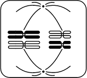
Determine the phase and type of division shown in the figure. Write down two numbers in the order indicated in the assignment, without separators (spaces, commas, etc.).
1) anaphase
2) metaphase
3) prophase
4) telophase
5) mitosis
6) meiosis I
7) meiosis II
Answer

Determine the phase and type of division shown in the figure. Write down two numbers in the order indicated in the assignment, without separators (spaces, commas, etc.).
1) anaphase
2) metaphase
3) prophase
4) telophase
5) mitosis
6) meiosis I
7) meiosis II
Answer
Determine the phase and type of division shown in the figure. Write down two numbers in the order indicated in the assignment, without separators (spaces, commas, etc.).
1) anaphase
2) metaphase
3) prophase
4) telophase
5) mitosis
6) meiosis I
7) meiosis II
Answer
© D.V. Pozdnyakov, 2009-2019
 New rules for personal property tax
 Features of the sale of an apartment with illegal redevelopment
 Rules and procedure for the exam